6EKV

Structure of OrfX2 from Clostridium botulinum A2


Experimental Data Snapshot

  • Method: X-RAY DIFFRACTION
  • Resolution: 2.10 Å
  • R-Value Free: 0.259 
  • R-Value Work: 0.208 
  • R-Value Observed: 0.211 

wwPDB Validation   3D Report Full Report


This is version 1.4 of the entry. See complete history


Literature

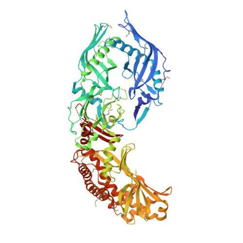
Crystal structures of OrfX2 and P47 from a Botulinum neurotoxin OrfX-type gene cluster.

Gustafsson, R.Berntsson, R.P.Martinez-Carranza, M.El Tekle, G.Odegrip, R.Johnson, E.A.Stenmark, P.

(2017) FEBS Lett 591: 3781-3792

  • DOI: https://doi.org/10.1002/1873-3468.12889
  • Primary Citation of Related Structures:  
    6EKT, 6EKV

  • PubMed Abstract: 

    Botulinum neurotoxins are highly toxic substances and are all encoded together with one of two alternative gene clusters, the HA or the OrfX gene cluster. Very little is known about the function and structure of the proteins encoded in the OrfX gene cluster, which in addition to the toxin contains five proteins (OrfX1, OrfX2, OrfX3, P47, and NTNH). We here present the structures of OrfX2 and P47, solved to 2.1 and 1.8 Å, respectively. We show that they belong to the TULIP protein superfamily, which are often involved in lipid binding. OrfX1 and OrfX2 were both found to bind phosphatidylinositol lipids.


  • Organizational Affiliation

    Department of Biochemistry and Biophysics, Stockholm University, Sweden.


Macromolecules
Find similar proteins by:  (by identity cutoff)  |  3D Structure
Entity ID: 1
MoleculeChains Sequence LengthOrganismDetailsImage
Toxin complex component ORF-X2750Clostridium botulinum A2 str. KyotoMutation(s): 0 
Gene Names: CLM_0892
UniProt
Find proteins for C1FUH4 (Clostridium botulinum (strain Kyoto / Type A2))
Explore C1FUH4 
Go to UniProtKB:  C1FUH4
Entity Groups  
Sequence Clusters30% Identity50% Identity70% Identity90% Identity95% Identity100% Identity
UniProt GroupC1FUH4
Sequence Annotations
Expand
  • Reference Sequence
Experimental Data & Validation

Experimental Data

  • Method: X-RAY DIFFRACTION
  • Resolution: 2.10 Å
  • R-Value Free: 0.259 
  • R-Value Work: 0.208 
  • R-Value Observed: 0.211 
  • Space Group: P 1 21 1
Unit Cell:
Length ( Å )Angle ( ˚ )
a = 39.646α = 90
b = 65.346β = 94.52
c = 161.455γ = 90
Software Package:
Software NamePurpose
REFMACrefinement
XDSdata reduction
Aimlessdata scaling
PHENIXphasing

Structure Validation

View Full Validation Report


Entry History & Funding Information

Deposition Data


Funding OrganizationLocationGrant Number
Swedish Research CouncilSweden2016-03599
European Molecular Biology OrganizationSwedenGA-2010- 267146
Swedish Research CouncilSweden2014-5667
Wenner-Gren FoundationSweden--
Swedish Cancer SocietySweden--

Revision History  (Full details and data files)

  • Version 1.0: 2017-10-25
    Type: Initial release
  • Version 1.1: 2017-11-08
    Changes: Database references
  • Version 1.2: 2017-12-06
    Changes: Database references, Structure summary
  • Version 1.3: 2018-01-24
    Changes: Source and taxonomy
  • Version 1.4: 2018-01-31
    Changes: Author supporting evidence