6EG0

Crystal structure of Dpr4 Ig1-Ig2 in complex with DIP-Eta Ig1-Ig3


Experimental Data Snapshot

  • Method: X-RAY DIFFRACTION
  • Resolution: 2.90 Å
  • R-Value Free: 0.264 
  • R-Value Work: 0.221 
  • R-Value Observed: 0.223 

wwPDB Validation   3D Report Full Report


Ligand Structure Quality Assessment 


This is version 2.1 of the entry. See complete history


Literature

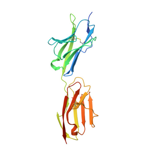
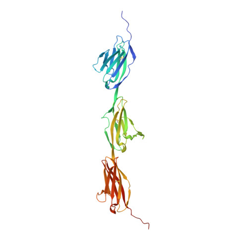
Neuron-Subtype-Specific Expression, Interaction Affinities, and Specificity Determinants of DIP/Dpr Cell Recognition Proteins.

Cosmanescu, F.Katsamba, P.S.Sergeeva, A.P.Ahlsen, G.Patel, S.D.Brewer, J.J.Tan, L.Xu, S.Xiao, Q.Nagarkar-Jaiswal, S.Nern, A.Bellen, H.J.Zipursky, S.L.Honig, B.Shapiro, L.

(2018) Neuron 100: 1385

  • DOI: https://doi.org/10.1016/j.neuron.2018.10.046
  • Primary Citation of Related Structures:  
    6EFY, 6EFZ, 6EG0, 6EG1

  • PubMed Abstract: 

    Binding between DIP and Dpr neuronal recognition proteins has been proposed to regulate synaptic connections between lamina and medulla neurons in the Drosophila visual system. Each lamina neuron was previously shown to express many Dprs. Here, we demonstrate, by contrast, that their synaptic partners typically express one or two DIPs, with binding specificities matched to the lamina neuron-expressed Dprs. A deeper understanding of the molecular logic of DIP/Dpr interaction requires quantitative studies on the properties of these proteins. We thus generated a quantitative affinity-based DIP/Dpr interactome for all DIP/Dpr protein family members. This revealed a broad range of affinities and identified homophilic binding for some DIPs and some Dprs. These data, along with full-length ectodomain DIP/Dpr and DIP/DIP crystal structures, led to the identification of molecular determinants of DIP/Dpr specificity. This structural knowledge, along with a comprehensive set of quantitative binding affinities, provides new tools for functional studies in vivo.


  • Organizational Affiliation

    Department of Biochemistry and Molecular Biophysics, Columbia University, New York, NY 10032, USA; Zuckerman Mind Brain Behavior Institute, Columbia University, New York, NY 10027, USA.


Macromolecules
Find similar proteins by:  (by identity cutoff)  |  3D Structure
Entity ID: 1
MoleculeChains Sequence LengthOrganismDetailsImage
Defective proboscis extension response 4220Drosophila melanogasterMutation(s): 0 
Gene Names: dpr4CG12593CG14707CT34497Dmel\CG33512Dpr-4Dpr4dpr4-RACG33512Dmel_CG33512
UniProt
Find proteins for Q59DX6 (Drosophila melanogaster)
Explore Q59DX6 
Go to UniProtKB:  Q59DX6
Entity Groups  
Sequence Clusters30% Identity50% Identity70% Identity90% Identity95% Identity100% Identity
UniProt GroupQ59DX6
Sequence Annotations
Expand
  • Reference Sequence
Find similar proteins by:  (by identity cutoff)  |  3D Structure
Entity ID: 2
MoleculeChains Sequence LengthOrganismDetailsImage
Dpr-interacting protein eta, isoform B314Drosophila melanogasterMutation(s): 0 
Gene Names: DIP-eta14010CT33567Dmel\CG14010CG14010Dmel_CG14010
UniProt
Find proteins for Q9VMN9 (Drosophila melanogaster)
Explore Q9VMN9 
Go to UniProtKB:  Q9VMN9
Entity Groups  
Sequence Clusters30% Identity50% Identity70% Identity90% Identity95% Identity100% Identity
UniProt GroupQ9VMN9
Sequence Annotations
Expand
  • Reference Sequence
Oligosaccharides

Help

Entity ID: 3
MoleculeChains Length2D Diagram Glycosylation3D Interactions
2-acetamido-2-deoxy-beta-D-glucopyranose-(1-4)-[alpha-L-fucopyranose-(1-6)]2-acetamido-2-deoxy-beta-D-glucopyranose
C
3N-Glycosylation
Glycosylation Resources
GlyTouCan:  G21290RB
GlyCosmos:  G21290RB
GlyGen:  G21290RB
Entity ID: 4
MoleculeChains Length2D Diagram Glycosylation3D Interactions
beta-D-mannopyranose-(1-4)-2-acetamido-2-deoxy-beta-D-glucopyranose-(1-4)-[alpha-L-fucopyranose-(1-6)]2-acetamido-2-deoxy-beta-D-glucopyranose
D
4N-Glycosylation
Glycosylation Resources
GlyTouCan:  G32152BH
GlyCosmos:  G32152BH
GlyGen:  G32152BH
Entity ID: 5
MoleculeChains Length2D Diagram Glycosylation3D Interactions
alpha-D-mannopyranose-(1-3)-beta-D-mannopyranose-(1-4)-2-acetamido-2-deoxy-beta-D-glucopyranose-(1-4)-[alpha-L-fucopyranose-(1-6)]2-acetamido-2-deoxy-beta-D-glucopyranose
E
5N-Glycosylation
Glycosylation Resources
GlyTouCan:  G42466VF
GlyCosmos:  G42466VF
GlyGen:  G42466VF
Entity ID: 6
MoleculeChains Length2D Diagram Glycosylation3D Interactions
2-acetamido-2-deoxy-beta-D-glucopyranose-(1-2)-alpha-D-mannopyranose-(1-3)-[2-acetamido-2-deoxy-beta-D-glucopyranose-(1-2)-alpha-D-mannopyranose-(1-6)]beta-D-mannopyranose-(1-4)-2-acetamido-2-deoxy-beta-D-glucopyranose-(1-4)-[alpha-L-fucopyranose-(1-6)]2-acetamido-2-deoxy-beta-D-glucopyranose
F
8N-Glycosylation
Glycosylation Resources
GlyTouCan:  G80858MF
GlyCosmos:  G80858MF
GlyGen:  G80858MF
Experimental Data & Validation

Experimental Data

  • Method: X-RAY DIFFRACTION
  • Resolution: 2.90 Å
  • R-Value Free: 0.264 
  • R-Value Work: 0.221 
  • R-Value Observed: 0.223 
  • Space Group: P 31 2 1
Unit Cell:
Length ( Å )Angle ( ˚ )
a = 94.294α = 90
b = 94.294β = 90
c = 212.463γ = 120
Software Package:
Software NamePurpose
PHENIXrefinement
XDSdata reduction
Aimlessdata scaling
PHASERphasing

Structure Validation

View Full Validation Report


Ligand Structure Quality Assessment 


Entry History & Funding Information

Deposition Data


Funding OrganizationLocationGrant Number
National Institutes of Health/National Institute of General Medical Sciences (NIH/NIGMS)United States--

Revision History  (Full details and data files)

  • Version 1.0: 2018-11-28
    Type: Initial release
  • Version 1.1: 2018-12-19
    Changes: Data collection, Database references
  • Version 1.2: 2019-01-02
    Changes: Data collection, Database references
  • Version 1.3: 2019-08-21
    Changes: Data collection, Structure summary
  • Version 1.4: 2020-01-01
    Changes: Author supporting evidence, Data collection
  • Version 2.0: 2020-07-29
    Type: Remediation
    Reason: Carbohydrate remediation
    Changes: Atomic model, Data collection, Derived calculations, Structure summary
  • Version 2.1: 2023-10-11
    Changes: Data collection, Database references, Derived calculations, Refinement description, Structure summary