6EF8

Cryo-EM of the OmcS nanowires from Geobacter sulfurreducens


Experimental Data Snapshot

  • Method: ELECTRON MICROSCOPY
  • Resolution: 3.70 Å
  • Aggregation State: FILAMENT 
  • Reconstruction Method: HELICAL 

wwPDB Validation   3D Report Full Report


This is version 1.2 of the entry. See complete history


Literature

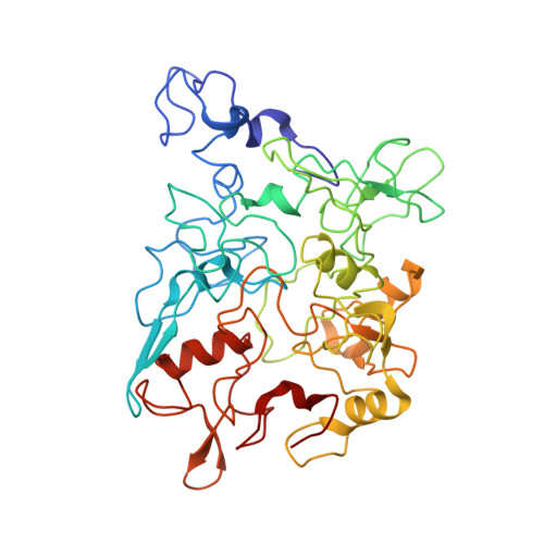
Structure of Microbial Nanowires Reveals Stacked Hemes that Transport Electrons over Micrometers.

Wang, F.Gu, Y.O'Brien, J.P.Yi, S.M.Yalcin, S.E.Srikanth, V.Shen, C.Vu, D.Ing, N.L.Hochbaum, A.I.Egelman, E.H.Malvankar, N.S.

(2019) Cell 177: 361-369.e10

  • DOI: https://doi.org/10.1016/j.cell.2019.03.029
  • Primary Citation of Related Structures:  
    6EF8

  • PubMed Abstract: 

    Long-range (>10 μm) transport of electrons along networks of Geobacter sulfurreducens protein filaments, known as microbial nanowires, has been invoked to explain a wide range of globally important redox phenomena. These nanowires were previously thought to be type IV pili composed of PilA protein. Here, we report a 3.7 Å resolution cryoelectron microscopy structure, which surprisingly reveals that, rather than PilA, G. sulfurreducens nanowires are assembled by micrometer-long polymerization of the hexaheme cytochrome OmcS, with hemes packed within ∼3.5-6 Å of each other. The inter-subunit interfaces show unique structural elements such as inter-subunit parallel-stacked hemes and axial coordination of heme by histidines from neighboring subunits. Wild-type OmcS filaments show 100-fold greater conductivity than other filaments from a ΔomcS strain, highlighting the importance of OmcS to conductivity in these nanowires. This structure explains the remarkable capacity of soil bacteria to transport electrons to remote electron acceptors for respiration and energy sharing.


  • Organizational Affiliation

    Department of Biochemistry and Molecular Genetics, University of Virginia School of Medicine, Charlottesville, VA 22908, USA.


Macromolecules
Find similar proteins by:  (by identity cutoff)  |  3D Structure
Entity ID: 1
MoleculeChains Sequence LengthOrganismDetailsImage
C-type cytochrome OmcS
A, B, C, D, E
A, B, C, D, E, F, G
407Geobacter sulfurreducens PCAMutation(s): 0 
UniProt
Find proteins for Q74A86 (Geobacter sulfurreducens (strain ATCC 51573 / DSM 12127 / PCA))
Explore Q74A86 
Go to UniProtKB:  Q74A86
Entity Groups  
Sequence Clusters30% Identity50% Identity70% Identity90% Identity95% Identity100% Identity
UniProt GroupQ74A86
Sequence Annotations
Expand
  • Reference Sequence
Small Molecules
Ligands 1 Unique
IDChains Name / Formula / InChI Key2D Diagram3D Interactions
HEC
Query on HEC

Download Ideal Coordinates CCD File 
AA [auth D]
BA [auth D]
CA [auth D]
DA [auth D]
EA [auth D]
AA [auth D],
BA [auth D],
CA [auth D],
DA [auth D],
EA [auth D],
FA [auth E],
GA [auth E],
H [auth A],
HA [auth E],
I [auth A],
IA [auth E],
J [auth A],
JA [auth E],
K [auth A],
KA [auth E],
L [auth A],
LA [auth F],
M [auth A],
MA [auth F],
N [auth B],
NA [auth F],
O [auth B],
OA [auth F],
P [auth B],
PA [auth F],
Q [auth B],
QA [auth F],
R [auth B],
RA [auth G],
S [auth B],
SA [auth G],
T [auth C],
TA [auth G],
U [auth C],
UA [auth G],
V [auth C],
VA [auth G],
W [auth C],
WA [auth G],
X [auth C],
Y [auth C],
Z [auth D]
HEME C
C34 H34 Fe N4 O4
HXQIYSLZKNYNMH-LJNAALQVSA-N
Experimental Data & Validation

Experimental Data

  • Method: ELECTRON MICROSCOPY
  • Resolution: 3.70 Å
  • Aggregation State: FILAMENT 
  • Reconstruction Method: HELICAL 
EM Software:
TaskSoftware PackageVersion
RECONSTRUCTIONSPIDER
MODEL REFINEMENTRosetta
MODEL REFINEMENTPHENIX
MODEL REFINEMENTCoot
MODEL REFINEMENTUCSF Chimera

Structure Validation

View Full Validation Report


Entry History & Funding Information

Deposition Data


Funding OrganizationLocationGrant Number
National Institutes of Health/National Human Genome Research Institute (NIH/NHGRI)United StatesR35GM122510
National Institutes of Health/National Human Genome Research Institute (NIH/NHGRI)United States1DP2AI138259-01
National Science Foundation (NSF, United States)United States1749662

Revision History  (Full details and data files)

  • Version 1.0: 2019-04-10
    Type: Initial release
  • Version 1.1: 2019-04-17
    Changes: Data collection, Database references
  • Version 1.2: 2019-11-27
    Changes: Author supporting evidence