6E4P

Structure of the T. brucei RRM domain in complex with RNA


Experimental Data Snapshot

  • Method: X-RAY DIFFRACTION
  • Resolution: 1.95 Å
  • R-Value Free: 0.229 
  • R-Value Work: 0.181 
  • R-Value Observed: 0.183 

wwPDB Validation   3D Report Full Report


This is version 1.3 of the entry. See complete history


Literature

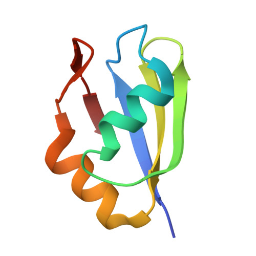
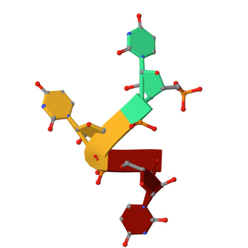
The RRM of the kRNA-editing protein TbRGG2 uses multiple surfaces to bind and remodel RNA.

Travis, B.Shaw, P.L.R.Liu, B.Ravindra, K.Iliff, H.Al-Hashimi, H.M.Schumacher, M.A.

(2019) Nucleic Acids Res 47: 2130-2142

  • DOI: https://doi.org/10.1093/nar/gky1259
  • Primary Citation of Related Structures:  
    6E4N, 6E4O, 6E4P

  • PubMed Abstract: 

    Kinetoplastid RNA (kRNA) editing takes place in the mitochondria of kinetoplastid protists and creates translatable mRNAs by uridine insertion/deletion. Extensively edited (pan-edited) transcripts contain quadruplex forming guanine stretches, which must be remodeled to promote uridine insertion/deletion. Here we show that the RRM domain of the essential kRNA-editing factor TbRGG2 binds poly(G) and poly(U) RNA and can unfold both. A region C-terminal to the RRM mediates TbRGG2 dimerization, enhancing RNA binding. A RRM-U4 RNA structure reveals a unique RNA-binding mechanism in which the two RRMs of the dimer employ aromatic residues outside the canonical RRM RNA-binding motifs to encase and wrench open the RNA, while backbone atoms specify the uridine bases. Notably, poly(G) RNA is bound via a different binding surface. Thus, these data indicate that TbRGG2 RRM can bind and remodel several RNA substrates suggesting how it might play multiple roles in the kRNA editing process.


  • Organizational Affiliation

    Department of Biochemistry, Duke University School of Medicine, Durham, NC 27710, USA.


Macromolecules

Find similar proteins by:  (by identity cutoff)  |  3D Structure
Entity ID: 2
MoleculeChains Sequence LengthOrganismDetailsImage
RNA-binding protein, putative71Trypanosoma bruceiMutation(s): 0 
UniProt
Find proteins for Q389P7 (Trypanosoma brucei brucei (strain 927/4 GUTat10.1))
Explore Q389P7 
Go to UniProtKB:  Q389P7
Entity Groups  
Sequence Clusters30% Identity50% Identity70% Identity90% Identity95% Identity100% Identity
UniProt GroupQ389P7
Sequence Annotations
Expand
  • Reference Sequence

Find similar nucleic acids by:  Sequence   |   3D Structure  

Entity ID: 1
MoleculeChains LengthOrganismImage
RNA (5'-R(P*UP*UP*UP*U)-3')A [auth J],
B [auth K]
4Trypanosoma brucei
Sequence Annotations
Expand
  • Reference Sequence
Experimental Data & Validation

Experimental Data

  • Method: X-RAY DIFFRACTION
  • Resolution: 1.95 Å
  • R-Value Free: 0.229 
  • R-Value Work: 0.181 
  • R-Value Observed: 0.183 
  • Space Group: P 1 21 1
Unit Cell:
Length ( Å )Angle ( ˚ )
a = 49.123α = 90
b = 126.297β = 118.57
c = 49.203γ = 90
Software Package:
Software NamePurpose
PHENIXrefinement
iMOSFLMdata reduction
SCALAdata scaling
MOLREPphasing

Structure Validation

View Full Validation Report


Entry History 

Deposition Data

Revision History  (Full details and data files)

  • Version 1.0: 2018-12-12
    Type: Initial release
  • Version 1.1: 2018-12-26
    Changes: Data collection, Database references
  • Version 1.2: 2019-03-13
    Changes: Data collection, Database references
  • Version 1.3: 2023-10-11
    Changes: Data collection, Database references, Refinement description