6E07

Crystal structure of Canton G6PD in complex with structural NADP


Experimental Data Snapshot

  • Method: X-RAY DIFFRACTION
  • Resolution: 2.60 Å
  • R-Value Free: 0.211 
  • R-Value Work: 0.187 
  • R-Value Observed: 0.189 

wwPDB Validation   3D Report Full Report


Ligand Structure Quality Assessment 


This is version 1.2 of the entry. See complete history


Literature

Correcting glucose-6-phosphate dehydrogenase deficiency with a small-molecule activator.

Hwang, S.Mruk, K.Rahighi, S.Raub, A.G.Chen, C.H.Dorn, L.E.Horikoshi, N.Wakatsuki, S.Chen, J.K.Mochly-Rosen, D.

(2018) Nat Commun 9: 4045-4045

  • DOI: https://doi.org/10.1038/s41467-018-06447-z
  • Primary Citation of Related Structures:  
    6E07, 6E08

  • PubMed Abstract: 

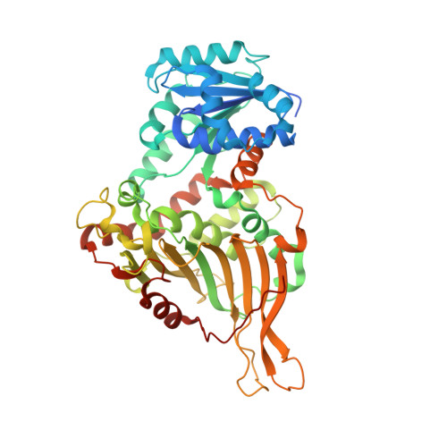
    Glucose-6-phosphate dehydrogenase (G6PD) deficiency, one of the most common human genetic enzymopathies, is caused by over 160 different point mutations and contributes to the severity of many acute and chronic diseases associated with oxidative stress, including hemolytic anemia and bilirubin-induced neurological damage particularly in newborns. As no medications are available to treat G6PD deficiency, here we seek to identify a small molecule that corrects it. Crystallographic study and mutagenesis analysis identify the structural and functional defect of one common mutant (Canton, R459L). Using high-throughput screening, we subsequently identify AG1, a small molecule that increases the activity of the wild-type, the Canton mutant and several other common G6PD mutants. AG1 reduces oxidative stress in cells and zebrafish. Furthermore, AG1 decreases chloroquine- or diamide-induced oxidative stress in human erythrocytes. Our study suggests that a pharmacological agent, of which AG1 may be a lead, will likely alleviate the challenges associated with G6PD deficiency.


  • Organizational Affiliation

    Department of Chemical and Systems Biology, Stanford University School of Medicine, Stanford, CA, 94305, USA.


Macromolecules
Find similar proteins by:  (by identity cutoff)  |  3D Structure
Entity ID: 1
MoleculeChains Sequence LengthOrganismDetailsImage
Glucose-6-phosphate 1-dehydrogenase515Homo sapiensMutation(s): 1 
Gene Names: G6PD
EC: 1.1.1.49
UniProt & NIH Common Fund Data Resources
Find proteins for P11413 (Homo sapiens)
Explore P11413 
Go to UniProtKB:  P11413
PHAROS:  P11413
GTEx:  ENSG00000160211 
Entity Groups  
Sequence Clusters30% Identity50% Identity70% Identity90% Identity95% Identity100% Identity
UniProt GroupP11413
Sequence Annotations
Expand
  • Reference Sequence
Small Molecules
Ligands 3 Unique
IDChains Name / Formula / InChI Key2D Diagram3D Interactions
NAP
Query on NAP

Download Ideal Coordinates CCD File 
EA [auth N]
I [auth L]
MA [auth Q]
QA [auth T]
R [auth C]
EA [auth N],
I [auth L],
MA [auth Q],
QA [auth T],
R [auth C],
UA [auth W],
X [auth F],
YA [auth B]
NADP NICOTINAMIDE-ADENINE-DINUCLEOTIDE PHOSPHATE
C21 H28 N7 O17 P3
XJLXINKUBYWONI-NNYOXOHSSA-N
PO4
Query on PO4

Download Ideal Coordinates CCD File 
FA [auth N]
J [auth L]
NA [auth Q]
RA [auth T]
S [auth C]
FA [auth N],
J [auth L],
NA [auth Q],
RA [auth T],
S [auth C],
VA [auth W],
Y [auth F],
ZA [auth B]
PHOSPHATE ION
O4 P
NBIIXXVUZAFLBC-UHFFFAOYSA-K
GOL
Query on GOL

Download Ideal Coordinates CCD File 
AA [auth F]
AB [auth B]
BA [auth F]
CA [auth F]
DA [auth F]
AA [auth F],
AB [auth B],
BA [auth F],
CA [auth F],
DA [auth F],
GA [auth N],
HA [auth N],
IA [auth N],
JA [auth N],
K [auth L],
KA [auth N],
L,
LA [auth N],
M [auth L],
N [auth L],
O [auth L],
OA [auth Q],
P [auth L],
PA [auth Q],
Q [auth L],
SA [auth T],
T [auth C],
TA [auth T],
U [auth C],
V [auth C],
W [auth C],
WA [auth W],
XA [auth W],
Z [auth F]
GLYCEROL
C3 H8 O3
PEDCQBHIVMGVHV-UHFFFAOYSA-N
Experimental Data & Validation

Experimental Data

  • Method: X-RAY DIFFRACTION
  • Resolution: 2.60 Å
  • R-Value Free: 0.211 
  • R-Value Work: 0.187 
  • R-Value Observed: 0.189 
  • Space Group: P 21 21 21
Unit Cell:
Length ( Å )Angle ( ˚ )
a = 127.12α = 90
b = 206.25β = 90
c = 211.84γ = 90
Software Package:
Software NamePurpose
iMOSFLMdata reduction
Aimlessdata scaling
MOLREPphasing
REFMACrefinement

Structure Validation

View Full Validation Report


Ligand Structure Quality Assessment 


Entry History 

Deposition Data

Revision History  (Full details and data files)

  • Version 1.0: 2018-07-25
    Type: Initial release
  • Version 1.1: 2018-10-17
    Changes: Data collection, Database references
  • Version 1.2: 2023-10-11
    Changes: Data collection, Database references, Refinement description