6DY3

Caenorhabditis elegans N-acylethanolamine-hydrolyzing acid amidase (NAAA) ortholog


Experimental Data Snapshot

  • Method: X-RAY DIFFRACTION
  • Resolution: 2.70 Å
  • R-Value Free: 0.215 
  • R-Value Work: 0.168 
  • R-Value Observed: 0.171 

wwPDB Validation   3D Report Full Report


This is version 2.1 of the entry. See complete history


Literature

Molecular mechanism of activation of the immunoregulatory amidase NAAA.

Gorelik, A.Gebai, A.Illes, K.Piomelli, D.Nagar, B.

(2018) Proc Natl Acad Sci U S A 115: E10032-E10040

  • DOI: https://doi.org/10.1073/pnas.1811759115
  • Primary Citation of Related Structures:  
    6DXW, 6DXX, 6DXY, 6DXZ, 6DY0, 6DY1, 6DY2, 6DY3

  • PubMed Abstract: 

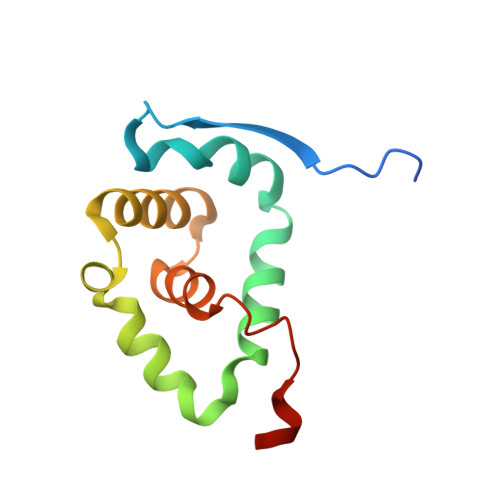
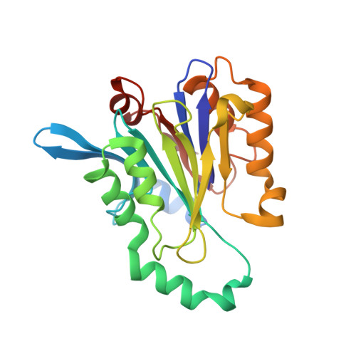
    Palmitoylethanolamide is a bioactive lipid that strongly alleviates pain and inflammation in animal models and in humans. Its signaling activity is terminated through degradation by N -acylethanolamine acid amidase (NAAA), a cysteine hydrolase expressed at high levels in immune cells. Pharmacological inhibitors of NAAA activity exert profound analgesic and antiinflammatory effects in rodent models, pointing to this protein as a potential target for therapeutic drug discovery. To facilitate these efforts and to better understand the molecular mechanism of action of NAAA, we determined crystal structures of this enzyme in various activation states and in complex with several ligands, including both a covalent and a reversible inhibitor. Self-proteolysis exposes the otherwise buried active site of NAAA to allow catalysis. Formation of a stable substrate- or inhibitor-binding site appears to be conformationally coupled to the interaction of a pair of hydrophobic helices in the enzyme with lipid membranes, resulting in the creation of a linear hydrophobic cavity near the active site that accommodates the ligand's acyl chain.


  • Organizational Affiliation

    Department of Biochemistry, McGill University, Montreal, H3G0B1, Canada.


Macromolecules
Find similar proteins by:  (by identity cutoff)  |  3D Structure
Entity ID: 1
MoleculeChains Sequence LengthOrganismDetailsImage
N-acylethanolamine-hydrolyzing acid amidase alpha-subunit
A, C, E, G
113Caenorhabditis elegansMutation(s): 0 
Gene Names: CELE_Y55D5A.3Y55D5A.3
EC: 3.5.1.60
UniProt
Find proteins for Q9GUI1 (Caenorhabditis elegans)
Explore Q9GUI1 
Go to UniProtKB:  Q9GUI1
Entity Groups  
Sequence Clusters30% Identity50% Identity70% Identity90% Identity95% Identity100% Identity
UniProt GroupQ9GUI1
Sequence Annotations
Expand
  • Reference Sequence
Find similar proteins by:  (by identity cutoff)  |  3D Structure
Entity ID: 2
MoleculeChains Sequence LengthOrganismDetailsImage
N-acylethanolamine-hydrolyzing acid amidase beta-subunit
B, D, F, H
234Caenorhabditis elegansMutation(s): 0 
Gene Names: CELE_Y55D5A.3Y55D5A.3
UniProt
Find proteins for Q9GUI1 (Caenorhabditis elegans)
Explore Q9GUI1 
Go to UniProtKB:  Q9GUI1
Entity Groups  
Sequence Clusters30% Identity50% Identity70% Identity90% Identity95% Identity100% Identity
UniProt GroupQ9GUI1
Sequence Annotations
Expand
  • Reference Sequence
Oligosaccharides

Help

Entity ID: 3
MoleculeChains Length2D Diagram Glycosylation3D Interactions
2-acetamido-2-deoxy-beta-D-glucopyranose-(1-4)-[alpha-L-fucopyranose-(1-6)]2-acetamido-2-deoxy-beta-D-glucopyranose
I, J
3N-Glycosylation
Glycosylation Resources
GlyTouCan:  G21290RB
GlyCosmos:  G21290RB
GlyGen:  G21290RB
Experimental Data & Validation

Experimental Data

  • Method: X-RAY DIFFRACTION
  • Resolution: 2.70 Å
  • R-Value Free: 0.215 
  • R-Value Work: 0.168 
  • R-Value Observed: 0.171 
  • Space Group: P 43
Unit Cell:
Length ( Å )Angle ( ˚ )
a = 89.614α = 90
b = 89.614β = 90
c = 207.058γ = 90
Software Package:
Software NamePurpose
PHENIXrefinement
HKL-2000data reduction
HKL-2000data scaling
PHASERphasing

Structure Validation

View Full Validation Report


Entry History & Funding Information

Deposition Data


Funding OrganizationLocationGrant Number
Canadian Institutes of Health Research (CIHR)CanadaMOP-133535

Revision History  (Full details and data files)

  • Version 1.0: 2018-09-26
    Type: Initial release
  • Version 1.1: 2018-10-10
    Changes: Data collection, Database references, Structure summary
  • Version 1.2: 2018-10-24
    Changes: Data collection, Database references, Structure summary
  • Version 1.3: 2018-11-07
    Changes: Data collection, Database references
  • Version 1.4: 2019-04-17
    Changes: Data collection, Structure summary
  • Version 1.5: 2020-01-08
    Changes: Author supporting evidence, Data collection
  • Version 2.0: 2020-07-29
    Type: Remediation
    Reason: Carbohydrate remediation
    Changes: Advisory, Atomic model, Data collection, Derived calculations, Structure summary
  • Version 2.1: 2023-10-11
    Changes: Data collection, Database references, Derived calculations, Refinement description, Structure summary