6DS5

Cryo EM structure of human SEIPIN


Experimental Data Snapshot

  • Method: ELECTRON MICROSCOPY
  • Resolution: 3.80 Å
  • Aggregation State: PARTICLE 
  • Reconstruction Method: SINGLE PARTICLE 

wwPDB Validation   3D Report Full Report


This is version 2.0 of the entry. See complete history


Literature

Human SEIPIN Binds Anionic Phospholipids.

Yan, R.Qian, H.Lukmantara, I.Gao, M.Du, X.Yan, N.Yang, H.

(2018) Dev Cell 47: 248-256.e4

  • DOI: https://doi.org/10.1016/j.devcel.2018.09.010
  • Primary Citation of Related Structures:  
    6DS5

  • PubMed Abstract: 

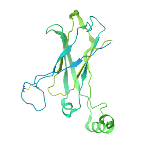
    The biogenesis of lipid droplets (LDs) and the development of adipocytes are two key aspects of mammalian fat storage. SEIPIN, an integral membrane protein of the endoplasmic reticulum (ER), plays a critical role in both LD formation and adipogenesis. The molecular function of SEIPIN, however, has yet to be elucidated. Here, we report the cryogenic electron microscopy structure of human SEIPIN at 3.8 Å resolution. SEIPIN exists as an undecamer, and this oligomerization state is critical for its physiological function. The evolutionarily conserved lumenal domain of SEIPIN forms an eight-stranded β sandwich fold. Both full-length SEIPIN and its lumenal domain can bind anionic phospholipids including phosphatidic acid. Our results suggest that SEIPIN forms a scaffold that helps maintain phospholipid homeostasis and surface tension of the ER.


  • Organizational Affiliation

    Tsinghua-Peking Joint Center for Life Sciences, Beijing Advanced Innovation Center for Structural Biology, State Key Laboratory of Membrane Biology, School of Life Sciences and School of Medicine, Tsinghua University, Beijing 100084, China.


Macromolecules
Find similar proteins by:  (by identity cutoff)  |  3D Structure
Entity ID: 1
MoleculeChains Sequence LengthOrganismDetailsImage
Seipin
A, B, C, D, E
A, B, C, D, E, F, G, H, I, J, K
409Homo sapiensMutation(s): 0 
Gene Names: BSCL2
Membrane Entity: Yes 
UniProt & NIH Common Fund Data Resources
Find proteins for Q96G97 (Homo sapiens)
Explore Q96G97 
Go to UniProtKB:  Q96G97
PHAROS:  Q96G97
GTEx:  ENSG00000168000 
Entity Groups  
Sequence Clusters30% Identity50% Identity70% Identity90% Identity95% Identity100% Identity
UniProt GroupQ96G97
Sequence Annotations
Expand
  • Reference Sequence
Oligosaccharides

Help

Entity ID: 2
MoleculeChains Length2D Diagram Glycosylation3D Interactions
2-acetamido-2-deoxy-beta-D-glucopyranose-(1-4)-2-acetamido-2-deoxy-beta-D-glucopyranose
L, M, N, O, P
L, M, N, O, P, Q, R, S, T, U, V
2N-Glycosylation
Glycosylation Resources
GlyTouCan:  G42666HT
GlyCosmos:  G42666HT
GlyGen:  G42666HT
Experimental Data & Validation

Experimental Data

  • Method: ELECTRON MICROSCOPY
  • Resolution: 3.80 Å
  • Aggregation State: PARTICLE 
  • Reconstruction Method: SINGLE PARTICLE 

Structure Validation

View Full Validation Report


Entry History & Funding Information

Deposition Data


Funding OrganizationLocationGrant Number
Ministry of Science and Technology (MoST, China)China2016YFA0502004
National Health and Medical Research Council (NHMRC, Australia)Australia1078117

Revision History  (Full details and data files)

  • Version 1.0: 2018-10-24
    Type: Initial release
  • Version 1.1: 2018-10-31
    Changes: Data collection, Database references
  • Version 1.2: 2019-12-18
    Changes: Author supporting evidence, Other
  • Version 2.0: 2020-07-29
    Type: Remediation
    Reason: Carbohydrate remediation
    Changes: Atomic model, Data collection, Derived calculations, Structure summary