Oligomeric Structure and Three-Dimensional Fold of the HIV gp41 Membrane-Proximal External Region and Transmembrane Domain in Phospholipid Bilayers.
Kwon, B., Lee, M., Waring, A.J., Hong, M.(2018) J Am Chem Soc 140: 8246-8259
- PubMed: 29888593
- DOI: https://doi.org/10.1021/jacs.8b04010
- Primary Citation of Related Structures:
6DLN - PubMed Abstract:
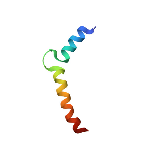
The HIV-1 glycoprotein, gp41, mediates fusion of the virus lipid envelope with the target cell membrane during virus entry into cells. Despite extensive studies of this protein, inconsistent and contradictory structural information abounds in the literature about the C-terminal membrane-interacting region of gp41. This C-terminal region contains the membrane-proximal external region (MPER), which harbors the epitopes for four broadly neutralizing antibodies, and the transmembrane domain (TMD), which anchors the protein to the virus lipid envelope. Due to the difficulty of crystallizing and solubilizing the MPER-TMD, most structural studies of this functionally important domain were carried out using truncated peptides either in the absence of membrane-mimetic solvents or bound to detergents and lipid bicelles. To determine the structural architecture of the MPER-TMD in the native environment of lipid membranes, we have now carried out a solid-state NMR study of the full MPER-TMD segment bound to cholesterol-containing phospholipid bilayers. 13 C chemical shifts indicate that the majority of the peptide is α-helical, except for the C-terminus of the TMD, which has moderate β-sheet character. Intermolecular 19 F- 19 F distance measurements of singly fluorinated peptides indicate that the MPER-TMD is trimerized in the virus-envelope mimetic lipid membrane. Intramolecular 13 C- 19 F distance measurements indicate the presence of a turn between the MPER helix and the TMD helix. This is supported by lipid-peptide and water-peptide 2D 1 H- 13 C correlation spectra, which indicate that the MPER binds to the membrane surface whereas the TMD spans the bilayer. Together, these data indicate that full-length MPER-TMD assembles into a trimeric helix-turn-helix structure in lipid membranes. We propose that the turn between the MPER and TMD may be important for inducing membrane defects in concert with negative-curvature lipid components such as cholesterol and phosphatidylethanolamine, while the surface-bound MPER helix may interact with N-terminal segments of the protein during late stages of membrane fusion.
Organizational Affiliation:
Department of Chemistry , Massachusetts Institute of Technology , 170 Albany Street , Cambridge , Massachusetts 02139 , United States.