6DET

The crystal structure of Tv2483 bound to L-arginine


Experimental Data Snapshot

  • Method: X-RAY DIFFRACTION
  • Resolution: 1.75 Å
  • R-Value Free: 0.199 
  • R-Value Work: 0.177 
  • R-Value Observed: 0.178 

wwPDB Validation   3D Report Full Report


Ligand Structure Quality Assessment 


This is version 1.2 of the entry. See complete history


Literature

Biophysical insights into a highly selective l-arginine-binding lipoprotein of a pathogenic treponeme.

Deka, R.K.Liu, W.Z.Tso, S.C.Norgard, M.V.Brautigam, C.A.

(2018) Protein Sci 27: 2037-2050

  • DOI: https://doi.org/10.1002/pro.3510
  • Primary Citation of Related Structures:  
    6DET

  • PubMed Abstract: 

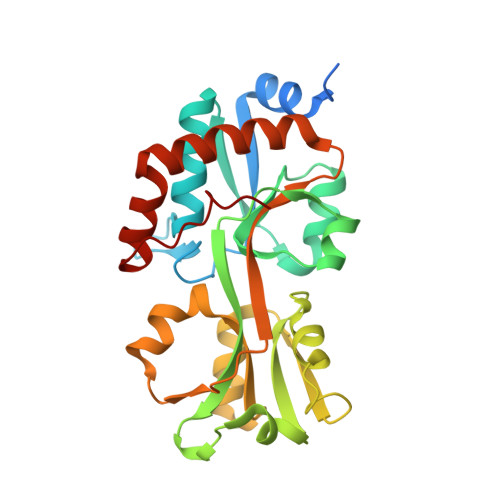
    Biophysical and biochemical studies on the lipoproteins and other periplasmic proteins from the spirochetal species Treponema pallidum have yielded numerous insights into the functioning of the organism's peculiar membrane organization, its nutritional requirements, and intermediary metabolism. However, not all T. pallidum proteins have proven to be amenable to biophysical studies. One such recalcitrant protein is Tp0309, a putative polar-amino-acid-binding protein of an ABC transporter system. To gain further information on its possible function, a homolog of the protein from the related species T. vincentii was used as a surrogate. This protein, Tv2483, was crystallized, resulting in the determination of its crystal structure at a resolution of 1.75 Å. The protein has a typical fold for a ligand-binding protein, and a single molecule of l-arginine was bound between its two lobes. Differential scanning fluorimetry and isothermal titration calorimetry experiments confirmed that l-arginine bound to the protein with unusually high selectivity. However, further comparison to Tp0309 showed differences in key amino-acid-binding residues may impart an alternate specificity for the T. pallidum protein.


  • Organizational Affiliation

    Departments of Microbiology, 5323 Harry Hines Blvd., Dallas, Texas, 75390.


Macromolecules
Find similar proteins by:  (by identity cutoff)  |  3D Structure
Entity ID: 1
MoleculeChains Sequence LengthOrganismDetailsImage
Tv2483266Treponema vincentiiMutation(s): 0 
UniProt
Find proteins for S3MDF0 (Treponema vincentii F0403)
Explore S3MDF0 
Go to UniProtKB:  S3MDF0
Entity Groups  
Sequence Clusters30% Identity50% Identity70% Identity90% Identity95% Identity100% Identity
UniProt GroupS3MDF0
Sequence Annotations
Expand
  • Reference Sequence
Small Molecules
Ligands 1 Unique
IDChains Name / Formula / InChI Key2D Diagram3D Interactions
ARG (Subject of Investigation/LOI)
Query on ARG

Download Ideal Coordinates CCD File 
B [auth A]ARGININE
C6 H15 N4 O2
ODKSFYDXXFIFQN-BYPYZUCNSA-O
Binding Affinity Annotations 
IDSourceBinding Affinity
ARG Binding MOAD:  6DET Kd: 6 (nM) from 1 assay(s)
Experimental Data & Validation

Experimental Data

  • Method: X-RAY DIFFRACTION
  • Resolution: 1.75 Å
  • R-Value Free: 0.199 
  • R-Value Work: 0.177 
  • R-Value Observed: 0.178 
  • Space Group: C 2 2 2
Unit Cell:
Length ( Å )Angle ( ˚ )
a = 104.586α = 90
b = 130.436β = 90
c = 48.991γ = 90
Software Package:
Software NamePurpose
PHENIXrefinement
HKL-3000data reduction
HKL-3000data scaling
PHASERphasing

Structure Validation

View Full Validation Report


Ligand Structure Quality Assessment 


Entry History & Funding Information

Deposition Data


Funding OrganizationLocationGrant Number
National Institutes of Health/National Institute Of Allergy and Infectious Diseases (NIH/NIAID)United StatesAI056305

Revision History  (Full details and data files)

  • Version 1.0: 2019-03-20
    Type: Initial release
  • Version 1.1: 2019-12-18
    Changes: Author supporting evidence
  • Version 1.2: 2024-03-13
    Changes: Data collection, Database references