6DE6

2.1 A resolution structure of histamine dehydrogenase from Rhizobium sp. 4-9


Experimental Data Snapshot

  • Method: X-RAY DIFFRACTION
  • Resolution: 2.10 Å
  • R-Value Free: 0.202 
  • R-Value Work: 0.164 
  • R-Value Observed: 0.166 

wwPDB Validation   3D Report Full Report


Ligand Structure Quality Assessment 


This is version 1.2 of the entry. See complete history


Literature

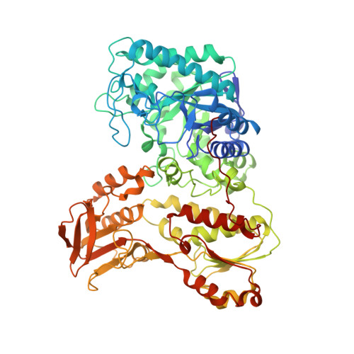
Structure of Rhizobium sp. 4-9 histamine dehydrogenase and analysis of the electron transfer pathway to an abiological electron acceptor.

Goyal, P.Deay 3rd, D.Seibold, S.Candido, A.C.L.Lovell, S.Battaile, K.P.Wilson, G.S.Richter, M.L.Petillo, P.A.

(2023) Arch Biochem Biophys : 109612-109612

  • DOI: https://doi.org/10.1016/j.abb.2023.109612
  • Primary Citation of Related Structures:  
    6DE6

  • PubMed Abstract: 

    Histamine dehydrogenase from the gram-negative bacterium Rhizobium sp. 4-9 (HaDHR) is a member of a small family of dehydrogenases containing a covalently attached FMN, and the only member so far identified to date that does not exhibit substrate inhibition. In this study, we present the 2.1 Å resolution crystal structure of HaDHR. This new structure allowed for the identification of the internal electron transfer pathway to abiological ferrocene-based mediators. Alanine 437 was identified as the exit point of electrons from the Fe 4 S 4 cluster. The enzyme was modified with a Ser436Cys mutation to facilitate covalent attachment of a ferrocene moiety. When modified with Fc-maleimide, this new construct demonstrated direct electron transfer from the enzyme to a gold electrode in a histamine concentration-dependent manner without the need for any additional electron mediators.


  • Organizational Affiliation

    Department of Molecular Biosciences, University of Kansas, Lawrence, KS, 66045, USA.


Macromolecules
Find similar proteins by:  (by identity cutoff)  |  3D Structure
Entity ID: 1
MoleculeChains Sequence LengthOrganismDetailsImage
Histamine dehydrogenase
A, B
696Rhizobium sp. 4-9Mutation(s): 0 
UniProt
Find proteins for Q60I59 (Rhizobium sp. 4-9)
Explore Q60I59 
Go to UniProtKB:  Q60I59
Entity Groups  
Sequence Clusters30% Identity50% Identity70% Identity90% Identity95% Identity100% Identity
UniProt GroupQ60I59
Sequence Annotations
Expand
  • Reference Sequence
Small Molecules
Ligands 4 Unique
IDChains Name / Formula / InChI Key2D Diagram3D Interactions
FMN
Query on FMN

Download Ideal Coordinates CCD File 
D [auth A],
G [auth B]
FLAVIN MONONUCLEOTIDE
C17 H21 N4 O9 P
FVTCRASFADXXNN-SCRDCRAPSA-N
ADP
Query on ADP

Download Ideal Coordinates CCD File 
C [auth A],
F [auth B]
ADENOSINE-5'-DIPHOSPHATE
C10 H15 N5 O10 P2
XTWYTFMLZFPYCI-KQYNXXCUSA-N
SF4
Query on SF4

Download Ideal Coordinates CCD File 
E [auth A],
H [auth B]
IRON/SULFUR CLUSTER
Fe4 S4
LJBDFODJNLIPKO-UHFFFAOYSA-N
PO4
Query on PO4

Download Ideal Coordinates CCD File 
I [auth B]PHOSPHATE ION
O4 P
NBIIXXVUZAFLBC-UHFFFAOYSA-K
Experimental Data & Validation

Experimental Data

  • Method: X-RAY DIFFRACTION
  • Resolution: 2.10 Å
  • R-Value Free: 0.202 
  • R-Value Work: 0.164 
  • R-Value Observed: 0.166 
  • Space Group: P 21 21 21
Unit Cell:
Length ( Å )Angle ( ˚ )
a = 103.223α = 90
b = 105.314β = 90
c = 140.038γ = 90
Software Package:
Software NamePurpose
XDSdata reduction
Aimlessdata scaling
PHASERphasing
PHENIXrefinement
PDB_EXTRACTdata extraction

Structure Validation

View Full Validation Report


Ligand Structure Quality Assessment 


Entry History 

Deposition Data

Revision History  (Full details and data files)

  • Version 1.0: 2019-05-15
    Type: Initial release
  • Version 1.1: 2023-05-17
    Changes: Database references, Derived calculations, Refinement description
  • Version 1.2: 2023-10-25
    Changes: Data collection, Refinement description