6DCH

Structure of isonitrile biosynthesis enzyme ScoE


Experimental Data Snapshot

  • Method: X-RAY DIFFRACTION
  • Resolution: 1.80 Å
  • R-Value Free: 0.207 
  • R-Value Work: 0.179 
  • R-Value Observed: 0.180 

wwPDB Validation   3D Report Full Report


This is version 1.3 of the entry. See complete history


Literature

Isonitrile Formation by a Non-Heme Iron(II)-Dependent Oxidase/Decarboxylase.

Harris, N.C.Born, D.A.Cai, W.Huang, Y.Martin, J.Khalaf, R.Drennan, C.L.Zhang, W.

(2018) Angew Chem Int Ed Engl 57: 9707-9710

  • DOI: https://doi.org/10.1002/anie.201804307
  • Primary Citation of Related Structures:  
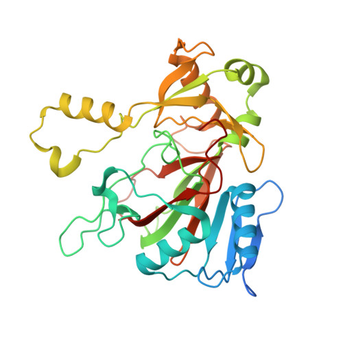
    6DCH

  • PubMed Abstract: 

    The electron-rich isonitrile is an important functionality in bioactive natural products, but its biosynthesis has been restricted to the IsnA family of isonitrile synthases. We herein provide the first structural and biochemical evidence of an alternative mechanism for isonitrile formation. ScoE, a putative non-heme iron(II)-dependent enzyme from Streptomyces coeruleorubidus, was shown to catalyze the conversion of (R)-3-((carboxymethyl)amino)butanoic acid to (R)-3-isocyanobutanoic acid through an oxidative decarboxylation mechanism. This work further provides a revised scheme for the biosynthesis of a unique class of isonitrile lipopeptides, of which several members are critical for the virulence of pathogenic mycobacteria.


  • Organizational Affiliation

    Department of Plant and Microbial Biology, University of California Berkeley, Berkeley, CA, 94720, USA.


Macromolecules
Find similar proteins by:  (by identity cutoff)  |  3D Structure
Entity ID: 1
MoleculeChains Sequence LengthOrganismDetailsImage
ScoE protein326Streptomyces coeruleorubidusMutation(s): 0 
Gene Names: ScoE
UniProt
Find proteins for A0A3B6UEU3 (Streptomyces coeruleorubidus)
Explore A0A3B6UEU3 
Go to UniProtKB:  A0A3B6UEU3
Entity Groups  
Sequence Clusters30% Identity50% Identity70% Identity90% Identity95% Identity100% Identity
UniProt GroupA0A3B6UEU3
Sequence Annotations
Expand
  • Reference Sequence
Experimental Data & Validation

Experimental Data

  • Method: X-RAY DIFFRACTION
  • Resolution: 1.80 Å
  • R-Value Free: 0.207 
  • R-Value Work: 0.179 
  • R-Value Observed: 0.180 
  • Space Group: P 43 21 2
Unit Cell:
Length ( Å )Angle ( ˚ )
a = 62.1α = 90
b = 62.1β = 90
c = 167.85γ = 90
Software Package:
Software NamePurpose
XDSdata reduction
XSCALEdata scaling
PHASERphasing
PHENIXrefinement
PDB_EXTRACTdata extraction

Structure Validation

View Full Validation Report


Entry History & Funding Information

Deposition Data


Funding OrganizationLocationGrant Number
Howard Hughes Medical Institute (HHMI)United States--

Revision History  (Full details and data files)

  • Version 1.0: 2018-06-27
    Type: Initial release
  • Version 1.1: 2018-08-29
    Changes: Data collection, Database references
  • Version 1.2: 2019-11-20
    Changes: Author supporting evidence
  • Version 1.3: 2023-10-11
    Changes: Data collection, Database references, Refinement description