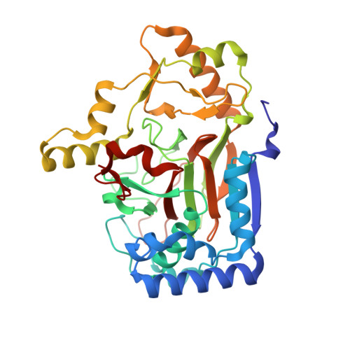
Two Distinct Mechanisms for C-C Desaturation by Iron(II)- and 2-(Oxo)glutarate-Dependent Oxygenases: Importance of alpha-Heteroatom Assistance.
Dunham, N.P., Chang, W.C., Mitchell, A.J., Martinie, R.J., Zhang, B., Bergman, J.A., Rajakovich, L.J., Wang, B., Silakov, A., Krebs, C., Boal, A.K., Bollinger, J.M.(2018) J Am Chem Soc 140: 7116-7126
- PubMed: 29708749
- DOI: https://doi.org/10.1021/jacs.8b01933
- Primary Citation of Related Structures:
6DAW, 6DAX, 6DAZ, 6DB2 - PubMed Abstract:
Hydroxylation of aliphatic carbons by nonheme Fe(IV)-oxo (ferryl) complexes proceeds by hydrogen-atom (H•) transfer (HAT) to the ferryl and subsequent coupling between the carbon radical and Fe(III)-coordinated oxygen (termed rebound). Enzymes that use H•-abstracting ferryl complexes for other transformations must either suppress rebound or further process hydroxylated intermediates. For olefin-installing C-C desaturations, it has been proposed that a second HAT to the Fe(III)-OH complex from the carbon α to the radical preempts rebound. Deuterium ( 2 H) at the second site should slow this step, potentially making rebound competitive. Desaturations mediated by two related l-arginine-modifying iron(II)- and 2-(oxo)glutarate-dependent (Fe/2OG) oxygenases behave oppositely in this key test, implicating different mechanisms. NapI, the l-Arg 4,5-desaturase from the naphthyridinomycin biosynthetic pathway, abstracts H• first from C5 but hydroxylates this site (leading to guanidine release) to the same modest extent whether C4 harbors 1 H or 2 H. By contrast, an unexpected 3,4-desaturation of l-homoarginine (l-hArg) by VioC, the l-Arg 3-hydroxylase from the viomycin biosynthetic pathway, is markedly disfavored relative to C4 hydroxylation when C3 (the second hydrogen donor) harbors 2 H. Anchimeric assistance by N6 permits removal of the C4-H as a proton in the NapI reaction, but, with no such assistance possible in the VioC desaturation, a second HAT step (from C3) is required. The close proximity (≤3.5 Å) of both l-hArg carbons to the oxygen ligand in an X-ray crystal structure of VioC harboring a vanadium-based ferryl mimic supports and rationalizes the sequential-HAT mechanism. The results suggest that, although the sequential-HAT mechanism is feasible, its geometric requirements may make competing hydroxylation unavoidable, thus explaining the presence of α-heteroatoms in nearly all native substrates for Fe/2OG desaturases.


