Entity ID: 1 |
|---|
| Molecule | Chains | Sequence Length | Organism | Details | Image |
|---|
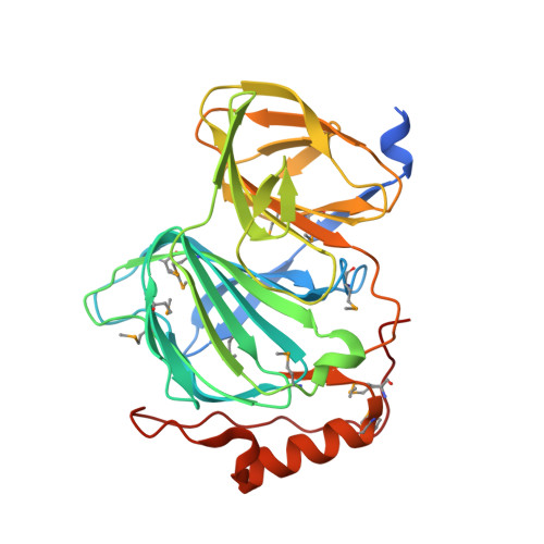
| Pirin family protein | | 318 | Acinetobacter baumannii | Mutation(s): 0
Gene Names: yhhW_2, yhhW_1, A388_00159, A7A65_05705, A7M90_08420, A7N09_08935, AZE33_00735, B4R90_09220, B9X91_00175, BGC29_04645, CAS83_08120, CBE85_14535, CEJ63_12860, CHQ89_16690, CPI82_15400, CV954_18565, LV38_01393
EC: 1.13.11.24
|  |
UniProt |
Find proteins for V5VHS3 (Acinetobacter baumannii) |
Entity Groups
|
| Sequence Clusters | 30% Identity50% Identity70% Identity90% Identity95% Identity100% Identity |
| UniProt Group | V5VHS3 |
Sequence Annotations |
|