6CYD

PDE2 in complex with compound 7


Experimental Data Snapshot

  • Method: X-RAY DIFFRACTION
  • Resolution: 1.69 Å
  • R-Value Free: 0.210 
  • R-Value Work: 0.182 
  • R-Value Observed: 0.184 

wwPDB Validation   3D Report Full Report


Ligand Structure Quality Assessment 


This is version 1.1 of the entry. See complete history


Literature

Structure-Guided Design and Procognitive Assessment of a Potent and Selective Phosphodiesterase 2A Inhibitor.

Stachel, S.J.Berger, R.Nomland, A.B.Ginnetti, A.T.Paone, D.V.Wang, D.Puri, V.Lange, H.Drott, J.Lu, J.Marcus, J.Dwyer, M.P.Suon, S.Uslaner, J.M.Smith, S.M.

(2018) ACS Med Chem Lett 9: 815-820

  • DOI: https://doi.org/10.1021/acsmedchemlett.8b00214
  • Primary Citation of Related Structures:  
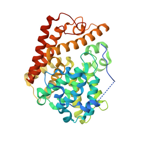
    6CYB, 6CYC, 6CYD

  • PubMed Abstract: 

    Herein we describe the development of a series of pyrazolopyrimidinone phosphodiesterase 2A (PDE2) inhibitors using structure-guided lead identification and design. The series was derived from informed chemotype replacement based on previously identified internal leads. The initially designed compound 3 , while potent on PDE2, displayed unsatisfactory selectivity against the other PDE2 isoforms. Compound 3 was subsequently optimized for improved PDE2 activity and isoform selectivity. Insights into the origins of PDE2 selectivity are described and verified using cocrystallography. An optimized lead, 4 , demonstrated improved performance in both a rodent and a nonhuman primate cognition model.


  • Organizational Affiliation

    Merck & Co. Inc., P.O. Box 4, West Point, Pennsylvania 19486, United States.


Macromolecules
Find similar proteins by:  (by identity cutoff)  |  3D Structure
Entity ID: 1
MoleculeChains Sequence LengthOrganismDetailsImage
cGMP-dependent 3',5'-cyclic phosphodiesterase
A, B
373Homo sapiensMutation(s): 0 
Gene Names: PDE2A
EC: 3.1.4.17
UniProt & NIH Common Fund Data Resources
Find proteins for O00408 (Homo sapiens)
Explore O00408 
Go to UniProtKB:  O00408
PHAROS:  O00408
GTEx:  ENSG00000186642 
Entity Groups  
Sequence Clusters30% Identity50% Identity70% Identity90% Identity95% Identity100% Identity
UniProt GroupO00408
Sequence Annotations
Expand
  • Reference Sequence
Small Molecules
Ligands 4 Unique
IDChains Name / Formula / InChI Key2D Diagram3D Interactions
FKG (Subject of Investigation/LOI)
Query on FKG

Download Ideal Coordinates CCD File 
E [auth A],
J [auth B]
3-(hydroxymethyl)-6-methyl-1-{(1S)-1-[4-(trifluoromethyl)phenyl]ethyl}-1,5-dihydro-4H-pyrazolo[3,4-d]pyrimidin-4-one
C16 H15 F3 N4 O2
WOPONWMJGIDXSF-QMMMGPOBSA-N
ZN
Query on ZN

Download Ideal Coordinates CCD File 
C [auth A],
H [auth B]
ZINC ION
Zn
PTFCDOFLOPIGGS-UHFFFAOYSA-N
EDO
Query on EDO

Download Ideal Coordinates CCD File 
F [auth A],
G [auth A],
K [auth B]
1,2-ETHANEDIOL
C2 H6 O2
LYCAIKOWRPUZTN-UHFFFAOYSA-N
MG
Query on MG

Download Ideal Coordinates CCD File 
D [auth A],
I [auth B]
MAGNESIUM ION
Mg
JLVVSXFLKOJNIY-UHFFFAOYSA-N
Binding Affinity Annotations 
IDSourceBinding Affinity
FKG Binding MOAD:  6CYD IC50: 2 (nM) from 1 assay(s)
Experimental Data & Validation

Experimental Data

  • Method: X-RAY DIFFRACTION
  • Resolution: 1.69 Å
  • R-Value Free: 0.210 
  • R-Value Work: 0.182 
  • R-Value Observed: 0.184 
  • Space Group: P 21 21 21
Unit Cell:
Length ( Å )Angle ( ˚ )
a = 71.94α = 90
b = 91.82β = 90
c = 102.83γ = 90
Software Package:
Software NamePurpose
BUSTERrefinement
XDSdata reduction
autoPROCdata scaling
REFMACphasing

Structure Validation

View Full Validation Report


Ligand Structure Quality Assessment 


Entry History 

Deposition Data

  • Released Date: 2018-09-19 
  • Deposition Author(s): Lu, J.

Revision History  (Full details and data files)

  • Version 1.0: 2018-09-19
    Type: Initial release
  • Version 1.1: 2024-03-06
    Changes: Data collection, Database references, Derived calculations, Refinement description