


Crystal structure of a dihydrofolate reductase from Mycobacterium smegmatis in complex with NADP and P218
Edwards, T.E., Abendroth, J., Lorimer, D.D., Horanyi, P.S., Seattle Structural Genomics Center for Infectious DiseaseTo be published.
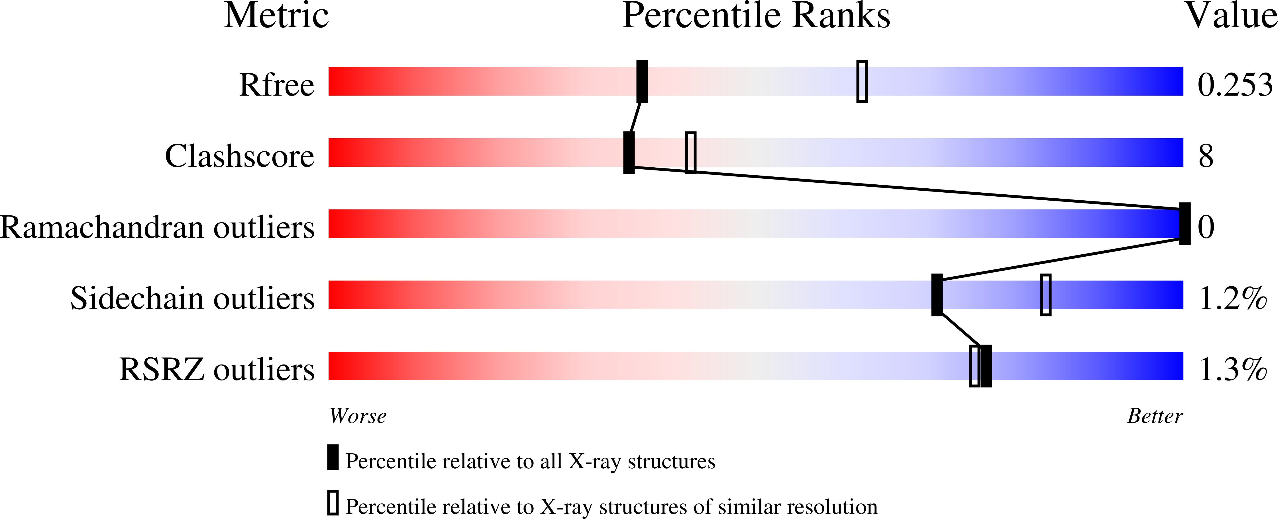
Experimental Data Snapshot
Entity ID: 1 | |||||
|---|---|---|---|---|---|
| Molecule | Chains | Sequence Length | Organism | Details | Image |
| Dihydrofolate reductase | 164 | Mycolicibacterium smegmatis MC2 155 | Mutation(s): 0 Gene Names: folA, MSMEI_2607 EC: 1.5.1.3 | 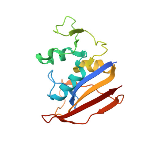 | |
UniProt | |||||
Find proteins for I7FC10 (Mycolicibacterium smegmatis (strain ATCC 700084 / mc(2)155)) Explore I7FC10 Go to UniProtKB: I7FC10 | |||||
Entity Groups | |||||
| Sequence Clusters | 30% Identity50% Identity70% Identity90% Identity95% Identity100% Identity | ||||
| UniProt Group | I7FC10 | ||||
Sequence AnnotationsExpand | |||||
| |||||
| Ligands 2 Unique | |||||
|---|---|---|---|---|---|
| ID | Chains | Name / Formula / InChI Key | 2D Diagram | 3D Interactions | |
| NAP Query on NAP | C [auth A], E [auth B] | NADP NICOTINAMIDE-ADENINE-DINUCLEOTIDE PHOSPHATE C21 H28 N7 O17 P3 XJLXINKUBYWONI-NNYOXOHSSA-N |  | ||
| MMV Query on MMV | D [auth A], F [auth B] | 3-(2-{3-[(2,4-diamino-6-ethylpyrimidin-5-yl)oxy]propoxy}phenyl)propanoic acid C18 H24 N4 O4 VDGXZSSDCDPCRF-UHFFFAOYSA-N |  | ||
| Length ( Å ) | Angle ( ˚ ) |
|---|---|
| a = 63.25 | α = 90 |
| b = 40.3 | β = 93.86 |
| c = 67.22 | γ = 90 |
| Software Name | Purpose |
|---|---|
| XDS | data reduction |
| XSCALE | data scaling |
| PHENIX | refinement |
| PDB_EXTRACT | data extraction |
| MOLREP | phasing |
RCSB PDB (citation) is hosted by
RCSB PDB is a member of the

