6CXH

Crystal structure of particulate methane monooxygenase from Methylomicrobium alcaliphilum 20Z


Experimental Data Snapshot

  • Method: X-RAY DIFFRACTION
  • Resolution: 2.70 Å
  • R-Value Free: 0.268 
  • R-Value Work: 0.213 
  • R-Value Observed: 0.216 

wwPDB Validation   3D Report Full Report


Ligand Structure Quality Assessment 


This is version 1.4 of the entry. See complete history


Literature

From micelles to bicelles: Effect of the membrane on particulate methane monooxygenase activity.

Ro, S.Y.Ross, M.O.Deng, Y.W.Batelu, S.Lawton, T.J.Hurley, J.D.Stemmler, T.L.Hoffman, B.M.Rosenzweig, A.C.

(2018) J Biol Chem 293: 10457-10465

  • DOI: https://doi.org/10.1074/jbc.RA118.003348
  • Primary Citation of Related Structures:  
    6CXH

  • PubMed Abstract: 

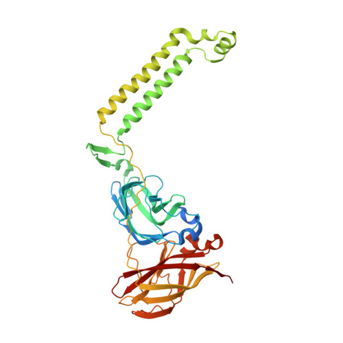
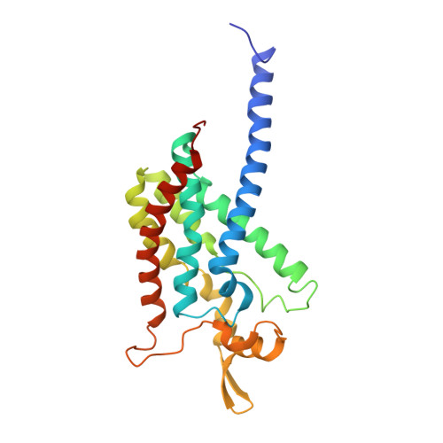
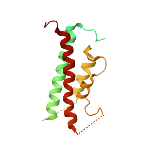
    Particulate methane monooxygenase (pMMO) is a copper-dependent integral membrane metalloenzyme that converts methane to methanol in methanotrophic bacteria. Studies of isolated pMMO have been hindered by loss of enzymatic activity upon its removal from the native membrane. To characterize pMMO in a membrane-like environment, we reconstituted pMMOs from Methylococcus ( Mcc. ) capsulatus (Bath) and Methylomicrobium ( Mm. ) alcaliphilum 20Z into bicelles. Reconstitution into bicelles recovers methane oxidation activity lost upon detergent solubilization and purification without substantial alterations to copper content or copper electronic structure, as observed by electron paramagnetic resonance (EPR) spectroscopy. These findings suggest that loss of pMMO activity upon isolation is due to removal from the membranes rather than caused by loss of the catalytic copper ions. A 2.7 Å resolution crystal structure of pMMO from Mm. alcaliphilum 20Z reveals a mononuclear copper center in the PmoB subunit and indicates that the transmembrane PmoC subunit may be conformationally flexible. Finally, results from extended X-ray absorption fine structure (EXAFS) analysis of pMMO from Mm. alcaliphilum 20Z were consistent with the observed monocopper center in the PmoB subunit. These results underscore the importance of studying membrane proteins in a membrane-like environment and provide valuable insight into pMMO function.


  • Organizational Affiliation

    From the Departments of Molecular Biosciences and Chemistry, Northwestern University, Evanston, Illinois 60208 and.


Macromolecules
Find similar proteins by:  (by identity cutoff)  |  3D Structure
Entity ID: 1
MoleculeChains Sequence LengthOrganismDetailsImage
Particulate methane monooxygenase, B subunit414Methylotuvimicrobium alcaliphilum 20ZMutation(s): 0 
EC: 1.14.13.25
Membrane Entity: Yes 
UniProt
Find proteins for G4SZ64 (Methylotuvimicrobium alcaliphilum (strain DSM 19304 / NCIMB 14124 / VKM B-2133 / 20Z))
Explore G4SZ64 
Go to UniProtKB:  G4SZ64
Entity Groups  
Sequence Clusters30% Identity50% Identity70% Identity90% Identity95% Identity100% Identity
UniProt GroupG4SZ64
Sequence Annotations
Expand
  • Reference Sequence
Find similar proteins by:  (by identity cutoff)  |  3D Structure
Entity ID: 2
MoleculeChains Sequence LengthOrganismDetailsImage
Particulate methane monooxygenase, A subunit247Methylotuvimicrobium alcaliphilum 20ZMutation(s): 0 
EC: 1.14.13.25
Membrane Entity: Yes 
UniProt
Find proteins for G4SZ63 (Methylotuvimicrobium alcaliphilum (strain DSM 19304 / NCIMB 14124 / VKM B-2133 / 20Z))
Explore G4SZ63 
Go to UniProtKB:  G4SZ63
Entity Groups  
Sequence Clusters30% Identity50% Identity70% Identity90% Identity95% Identity100% Identity
UniProt GroupG4SZ63
Sequence Annotations
Expand
  • Reference Sequence
Find similar proteins by:  (by identity cutoff)  |  3D Structure
Entity ID: 3
MoleculeChains Sequence LengthOrganismDetailsImage
Particulate methane monooxygenase, C subunit250Methylotuvimicrobium alcaliphilum 20ZMutation(s): 0 
EC: 1.14.13.25
Membrane Entity: Yes 
UniProt
Find proteins for G4SZ62 (Methylotuvimicrobium alcaliphilum (strain DSM 19304 / NCIMB 14124 / VKM B-2133 / 20Z))
Explore G4SZ62 
Go to UniProtKB:  G4SZ62
Entity Groups  
Sequence Clusters30% Identity50% Identity70% Identity90% Identity95% Identity100% Identity
UniProt GroupG4SZ62
Sequence Annotations
Expand
  • Reference Sequence
Experimental Data & Validation

Experimental Data

  • Method: X-RAY DIFFRACTION
  • Resolution: 2.70 Å
  • R-Value Free: 0.268 
  • R-Value Work: 0.213 
  • R-Value Observed: 0.216 
  • Space Group: P 63
Unit Cell:
Length ( Å )Angle ( ˚ )
a = 143.84α = 90
b = 143.84β = 90
c = 146.152γ = 120
Software Package:
Software NamePurpose
PHENIXrefinement
HKL-2000data reduction
HKL-2000data scaling

Structure Validation

View Full Validation Report


Ligand Structure Quality Assessment 


Entry History & Funding Information

Deposition Data


Funding OrganizationLocationGrant Number
National Institutes of Health/National Human Genome Research Institute (NIH/NHGRI)United StatesGM118035
National Institutes of Health/National Human Genome Research Institute (NIH/NHGRI)United States5T32GM008382

Revision History  (Full details and data files)

  • Version 1.0: 2018-05-16
    Type: Initial release
  • Version 1.1: 2018-05-23
    Changes: Data collection, Database references
  • Version 1.2: 2018-07-18
    Changes: Data collection, Database references
  • Version 1.3: 2019-12-18
    Changes: Author supporting evidence
  • Version 1.4: 2023-10-04
    Changes: Data collection, Database references, Refinement description