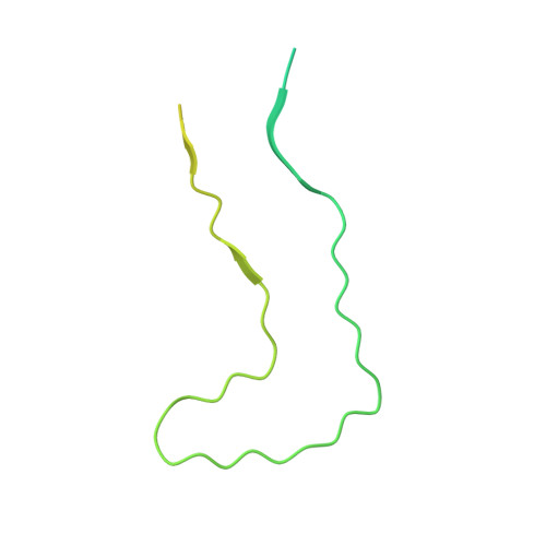
6CU8

Alpha Synuclein fibril formed by full length protein - Twister Polymorph


Experimental Data Snapshot

  • Method: ELECTRON MICROSCOPY
  • Resolution: 3.60 Å
  • Aggregation State: FILAMENT 
  • Reconstruction Method: HELICAL 

wwPDB Validation   3D Report Full Report


This is version 1.5 of the entry. See complete history


Literature

Cryo-EM of full-length alpha-synuclein reveals fibril polymorphs with a common structural kernel.

Li, B.Ge, P.Murray, K.A.Sheth, P.Zhang, M.Nair, G.Sawaya, M.R.Shin, W.S.Boyer, D.R.Ye, S.Eisenberg, D.S.Zhou, Z.H.Jiang, L.

(2018) Nat Commun 9: 3609-3609

  • DOI: https://doi.org/10.1038/s41467-018-05971-2
  • Primary Citation of Related Structures:  
    6CU7, 6CU8

  • PubMed Abstract: 

    α-Synuclein (aSyn) fibrillar polymorphs have distinct in vitro and in vivo seeding activities, contributing differently to synucleinopathies. Despite numerous prior attempts, how polymorphic aSyn fibrils differ in atomic structure remains elusive. Here, we present fibril polymorphs from the full-length recombinant human aSyn and their seeding capacity and cytotoxicity in vitro. By cryo-electron microscopy helical reconstruction, we determine the structures of the two predominant species, a rod and a twister, both at 3.7 Å resolution. Our atomic models reveal that both polymorphs share a kernel structure of a bent β-arch, but differ in their inter-protofilament interfaces. Thus, different packing of the same kernel structure gives rise to distinct fibril polymorphs. Analyses of disease-related familial mutations suggest their potential contribution to the pathogenesis of synucleinopathies by altering population distribution of the fibril polymorphs. Drug design targeting amyloid fibrils in neurodegenerative diseases should consider the formation and distribution of concurrent fibril polymorphs.


  • Organizational Affiliation

    Department of Neurology, David Geffen School of Medicine, UCLA, Los Angeles, CA, 90095, USA.


Macromolecules
Find similar proteins by:  (by identity cutoff)  |  3D Structure
Entity ID: 1
MoleculeChains Sequence LengthOrganismDetailsImage
Alpha-synuclein140Homo sapiensMutation(s): 0 
Gene Names: SNCANACPPARK1
UniProt & NIH Common Fund Data Resources
Find proteins for P37840 (Homo sapiens)
Explore P37840 
Go to UniProtKB:  P37840
PHAROS:  P37840
GTEx:  ENSG00000145335 
Entity Groups  
Sequence Clusters30% Identity50% Identity70% Identity90% Identity95% Identity100% Identity
UniProt GroupP37840
Sequence Annotations
Expand
  • Reference Sequence
Experimental Data & Validation

Experimental Data

  • Method: ELECTRON MICROSCOPY
  • Resolution: 3.60 Å
  • Aggregation State: FILAMENT 
  • Reconstruction Method: HELICAL 
EM Software:
TaskSoftware PackageVersion
MODEL REFINEMENTCNS1.21
MODEL REFINEMENTPHENIX1.10.1-2155
RECONSTRUCTIONRELION2.0

Structure Validation

View Full Validation Report


Entry History & Funding Information

Deposition Data


Funding OrganizationLocationGrant Number
XSEDEUnited StatesMCB140140
National Institutes of Health/National Institute Of Allergy and Infectious Diseases (NIH/NIAID)United States1R01AI094386
National Institutes of Health/National Institute of General Medical Sciences (NIH/NIGMS)United States1R01GM071940

Revision History  (Full details and data files)

  • Version 1.0: 2018-09-12
    Type: Initial release
  • Version 1.1: 2018-09-19
    Changes: Data collection, Database references
  • Version 1.2: 2019-02-27
    Changes: Data collection, Database references
  • Version 1.3: 2019-11-06
    Changes: Author supporting evidence, Data collection
  • Version 1.4: 2019-12-18
    Changes: Author supporting evidence, Other
  • Version 1.5: 2024-03-13
    Changes: Data collection, Database references