6CSL

Pneumococcal PhtD protein 269-339 fragment with bound Zn(II)


Experimental Data Snapshot

  • Method: X-RAY DIFFRACTION
  • Resolution: 1.92 Å
  • R-Value Free: 0.198 
  • R-Value Work: 0.148 
  • R-Value Observed: 0.153 

wwPDB Validation   3D Report Full Report


This is version 1.3 of the entry. See complete history


Literature

Structural characterisation of the HT3 motif of the polyhistidine triad protein D from Streptococcus pneumoniae.

Luo, Z.Pederick, V.G.Paton, J.C.McDevitt, C.A.Kobe, B.

(2018) FEBS Lett 592: 2341-2350

  • DOI: https://doi.org/10.1002/1873-3468.13122
  • Primary Citation of Related Structures:  
    6CSL

  • PubMed Abstract: 

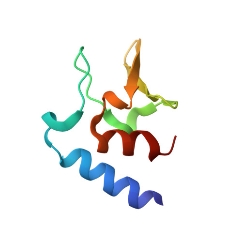
    The bacterium Streptococcus pneumoniae (the pneumococcus) is a major human pathogen that requires Zn 2+ for its survival and virulence in the host environment. Polyhistidine triad protein D (PhtD) has a known role in pneumococcal Zn 2+ homeostasis. However, the mechanistic basis of PhtD function remains unclear, partly due to a lack of structural information. Here, we determined the crystal structure of the fragment PhtD 269-339 , containing the third Zn 2+ -binding histidine triad (HT) motif of the protein. Analysis of the structure suggests that Zn 2+ binding occurs at the surface of the protein and that all five HT motifs in the protein bind Zn 2+ and share similar structures. These new structural insights aid in our understanding of how the Pht proteins facilitate pneumococcal Zn 2+ acquisition.


  • Organizational Affiliation

    School of Chemistry and Molecular Biosciences, University of Queensland, Brisbane, Queensland, Australia.


Macromolecules
Find similar proteins by:  (by identity cutoff)  |  3D Structure
Entity ID: 1
MoleculeChains Sequence LengthOrganismDetailsImage
Histidine triad protein D71Streptococcus pneumoniaeMutation(s): 0 
Gene Names: phtD
UniProt
Find proteins for Q8DQ08 (Streptococcus pneumoniae (strain ATCC BAA-255 / R6))
Explore Q8DQ08 
Go to UniProtKB:  Q8DQ08
Entity Groups  
Sequence Clusters30% Identity50% Identity70% Identity90% Identity95% Identity100% Identity
UniProt GroupQ8DQ08
Sequence Annotations
Expand
  • Reference Sequence
Small Molecules
Ligands 1 Unique
IDChains Name / Formula / InChI Key2D Diagram3D Interactions
ZN
Query on ZN

Download Ideal Coordinates CCD File 
B [auth A]ZINC ION
Zn
PTFCDOFLOPIGGS-UHFFFAOYSA-N
Experimental Data & Validation

Experimental Data

  • Method: X-RAY DIFFRACTION
  • Resolution: 1.92 Å
  • R-Value Free: 0.198 
  • R-Value Work: 0.148 
  • R-Value Observed: 0.153 
  • Space Group: C 1 2 1
Unit Cell:
Length ( Å )Angle ( ˚ )
a = 59.034α = 90
b = 47.978β = 107.13
c = 23.595γ = 90
Software Package:
Software NamePurpose
PHENIXrefinement
Aimlessdata scaling
PDB_EXTRACTdata extraction
XDSdata reduction
AutoSolphasing

Structure Validation

View Full Validation Report


Entry History & Funding Information

Deposition Data


Funding OrganizationLocationGrant Number
National Health and Medical Research Council (NHMRC, Australia)Australia1071659
National Health and Medical Research Council (NHMRC, Australia)Australia1080784
National Health and Medical Research Council (NHMRC, Australia)Australia1122582
Australian Research Council (ARC)AustraliaDP150104515
Australian Research Council (ARC)AustraliaDP170102102

Revision History  (Full details and data files)

  • Version 1.0: 2018-06-13
    Type: Initial release
  • Version 1.1: 2018-09-12
    Changes: Data collection, Database references
  • Version 1.2: 2020-01-01
    Changes: Author supporting evidence
  • Version 1.3: 2024-03-13
    Changes: Data collection, Database references