6CSJ

Structure of a Bacillus coagulans polyol dehydrogenase double mutant with an acquired D-lactate dehydrogenase activity


Experimental Data Snapshot

  • Method: X-RAY DIFFRACTION
  • Resolution: 2.40 Å
  • R-Value Free: 0.269 
  • R-Value Work: 0.237 

wwPDB Validation   3D Report Full Report


This is version 1.3 of the entry. See complete history


Literature

Kinetic characterization and structure analysis of an altered polyol dehydrogenase with d-lactate dehydrogenase activity.

Chauliac, D.Wang, Q.St John, F.J.Jones, G.Hurlbert, J.C.Ingram, L.O.Shanmugam, K.T.

(2020) Protein Sci 29: 2387-2397

  • DOI: https://doi.org/10.1002/pro.3963
  • Primary Citation of Related Structures:  
    6CSJ

  • PubMed Abstract: 

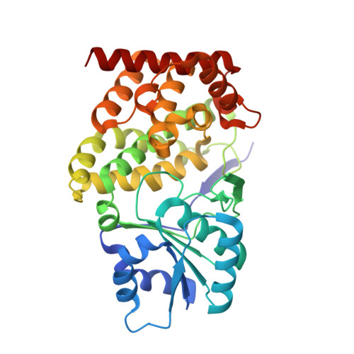
    During adaptive metabolic evolution a native glycerol dehydrogenase (GDH) acquired a d-lactate dehydrogenase (LDH) activity. Two active-site amino acid changes were detected in the altered protein. Biochemical studies along with comparative structure analysis using an X-ray crystallographic structure model of the protein with the two different amino acids allowed prediction of pyruvate binding into the active site. We propose that the F245S alteration increased the capacity of the glycerol binding site and facilitated hydrogen bonding between the S245 γ-O and the C1 carboxylate of pyruvate. To our knowledge, this is the first GDH to gain LDH activity due to an active site amino acid change, a desired result of in vivo enzyme evolution.


  • Organizational Affiliation

    Department of Microbiology and Cell Science, University of Florida, Gainesville, Florida, USA.


Macromolecules
Find similar proteins by:  (by identity cutoff)  |  3D Structure
Entity ID: 1
MoleculeChains Sequence LengthOrganismDetailsImage
Glycerol dehydrogenase
A, B, C, D, E
A, B, C, D, E, F, G, H
366Heyndrickxia coagulansMutation(s): 2 
Gene Names: ldhA
EC: 1.1.1.6
UniProt
Find proteins for A0A150JSL8 (Heyndrickxia coagulans)
Explore A0A150JSL8 
Go to UniProtKB:  A0A150JSL8
Entity Groups  
Sequence Clusters30% Identity50% Identity70% Identity90% Identity95% Identity100% Identity
UniProt GroupA0A150JSL8
Sequence Annotations
Expand
  • Reference Sequence
Experimental Data & Validation

Experimental Data

  • Method: X-RAY DIFFRACTION
  • Resolution: 2.40 Å
  • R-Value Free: 0.269 
  • R-Value Work: 0.237 
  • Space Group: C 2 2 21
Unit Cell:
Length ( Å )Angle ( ˚ )
a = 211.672α = 90
b = 211.772β = 90
c = 149.43γ = 90
Software Package:
Software NamePurpose
REFMACrefinement
PDB_EXTRACTdata extraction
HKL-3000data reduction
HKL-3000data scaling
MOLREPphasing

Structure Validation

View Full Validation Report


Entry History & Funding Information

Deposition Data


Funding OrganizationLocationGrant Number
National Institutes of Health/National Center for Research Resources (NIH/NCRR)United States5 P20 RR16461

Revision History  (Full details and data files)

  • Version 1.0: 2019-07-03
    Type: Initial release
  • Version 1.1: 2019-12-04
    Changes: Author supporting evidence
  • Version 1.2: 2021-01-13
    Changes: Database references
  • Version 1.3: 2023-10-04
    Changes: Data collection, Database references, Refinement description