6CRM

Crystal Structure of RecQ catalytic core from C. sakazakii bound to an unfolded G-quadruplex


Experimental Data Snapshot

  • Method: X-RAY DIFFRACTION
  • Resolution: 2.19 Å
  • R-Value Free: 0.240 
  • R-Value Work: 0.205 
  • R-Value Observed: 0.207 

wwPDB Validation   3D Report Full Report


This is version 1.4 of the entry. See complete history


Literature

A guanine-flipping and sequestration mechanism for G-quadruplex unwinding by RecQ helicases.

Voter, A.F.Qiu, Y.Tippana, R.Myong, S.Keck, J.L.

(2018) Nat Commun 9: 4201-4201

  • DOI: https://doi.org/10.1038/s41467-018-06751-8
  • Primary Citation of Related Structures:  
    6CRM

  • PubMed Abstract: 

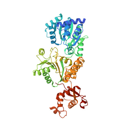
    Homeostatic regulation of G-quadruplexes (G4s), four-stranded structures that can form in guanine-rich nucleic acids, requires G4 unwinding helicases. The mechanisms that mediate G4 unwinding remain unknown. We report the structure of a bacterial RecQ DNA helicase bound to resolved G4 DNA. Unexpectedly, a guanine base from the unwound G4 is sequestered within a guanine-specific binding pocket. Disruption of the pocket in RecQ blocks G4 unwinding, but not G4 binding or duplex DNA unwinding, indicating its essential role in structure-specific G4 resolution. A novel guanine-flipping and sequestration model that may be applicable to other G4-resolving helicases emerges from these studies.


  • Organizational Affiliation

    Department of Biomolecular Chemistry, University of Wisconsin School of Medicine and Public Health, Madison, WI, 53706, USA.


Macromolecules

Find similar proteins by:  (by identity cutoff)  |  3D Structure
Entity ID: 1
MoleculeChains Sequence LengthOrganismDetailsImage
RecQ541Cronobacter sakazakii ATCC BAA-894Mutation(s): 0 
Gene Names: ESA_03738
UniProt
Find proteins for A7MQK9 (Cronobacter sakazakii (strain ATCC BAA-894))
Explore A7MQK9 
Go to UniProtKB:  A7MQK9
Entity Groups  
Sequence Clusters30% Identity50% Identity70% Identity90% Identity95% Identity100% Identity
UniProt GroupA7MQK9
Sequence Annotations
Expand
  • Reference Sequence
Find similar nucleic acids by:  (by identity cutoff)  |  3D Structure
Entity ID: 2
MoleculeChains LengthOrganismImage
DNA (5'-D(P*GP*GP*GP*TP*CP*GP*GP*TP*GP*CP*CP*TP*TP*A)-3')37synthetic construct
Sequence Annotations
Expand
  • Reference Sequence
Small Molecules
Ligands 1 Unique
IDChains Name / Formula / InChI Key2D Diagram3D Interactions
ZN
Query on ZN

Download Ideal Coordinates CCD File 
C [auth A]ZINC ION
Zn
PTFCDOFLOPIGGS-UHFFFAOYSA-N
Experimental Data & Validation

Experimental Data

  • Method: X-RAY DIFFRACTION
  • Resolution: 2.19 Å
  • R-Value Free: 0.240 
  • R-Value Work: 0.205 
  • R-Value Observed: 0.207 
  • Space Group: P 21 21 2
Unit Cell:
Length ( Å )Angle ( ˚ )
a = 78.468α = 90
b = 94.67β = 90
c = 98.906γ = 90
Software Package:
Software NamePurpose
PHENIXrefinement
XDSdata scaling
PHENIXphasing
PHENIXmodel building
XDSdata reduction

Structure Validation

View Full Validation Report


Entry History & Funding Information

Deposition Data


Funding OrganizationLocationGrant Number
National Institutes of Health/National Institute of General Medical Sciences (NIH/NIGMS)United StatesR01 GM098885

Revision History  (Full details and data files)

  • Version 1.0: 2018-10-17
    Type: Initial release
  • Version 1.1: 2018-10-24
    Changes: Data collection, Database references
  • Version 1.2: 2019-02-20
    Changes: Author supporting evidence, Data collection
  • Version 1.3: 2020-01-01
    Changes: Author supporting evidence
  • Version 1.4: 2023-10-04
    Changes: Data collection, Database references, Refinement description