6CPU

Crystal structure of yeast caPDE2

  • Classification: HYDROLASE
  • Organism(s): Candida albicans
  • Expression System: Escherichia coli BL21
  • Mutation(s): No 

  • Deposited: 2018-03-14 Released: 2019-02-20 
  • Deposition Author(s): Ke, H., Wang, y.
  • Funding Organization(s): National Institutes of Health/National Institute of General Medical Sciences (NIH/NIGMS)

Experimental Data Snapshot

  • Method: X-RAY DIFFRACTION
  • Resolution: 1.80 Å
  • R-Value Free: 0.226 
  • R-Value Work: 0.199 
  • R-Value Observed: 0.201 

wwPDB Validation   3D Report Full Report


This is version 1.5 of the entry. See complete history


Literature

Crystal Structures of Candida albicans Phosphodiesterase 2 and Implications for Its Biological Functions.

Yao, T.Huang, Y.Zhang, M.Chen, Y.Pei, H.Shi, J.Wang, H.Wang, Y.Ke, H.

(2018) Biochemistry 57: 6070-6077

  • DOI: https://doi.org/10.1021/acs.biochem.8b00707
  • Primary Citation of Related Structures:  
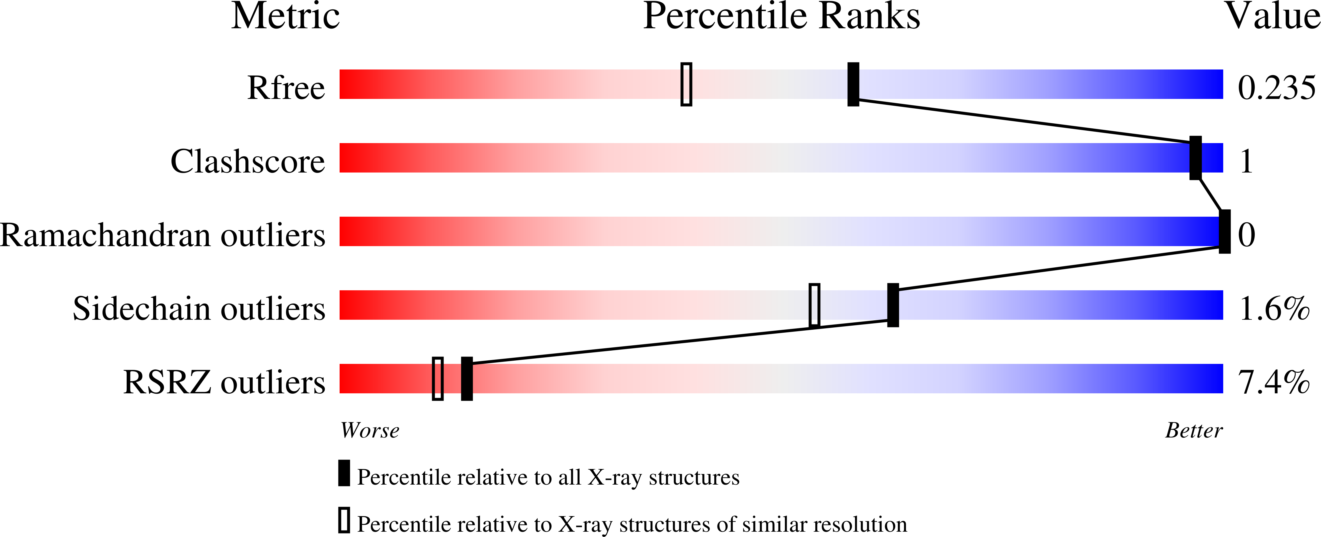
    6CPT, 6CPU

  • PubMed Abstract: 

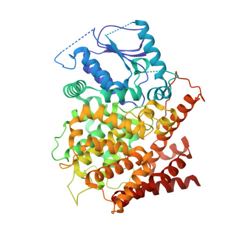
    The cAMP signaling system plays important roles in the physiological processes of pathogen yeast Candida albicans, but its functional mechanism has not been well illustrated. Here, we report the enzymatic characterization and crystal structures of C. albicans phosphodiesterase 2 (caPDE2) in the unliganded and 3-isobutyl-1-methylxanthine-complexed forms. caPDE2 is a monomer in liquid and crystal states and specifically hydrolyzes cAMP with a K M of 35 nM. It does not effectively hydrolyze cGMP as shown by the 1.32 × 10 5 -fold specificity of cAMP/cGMP. The crystal structure of caPDE2 shows significant differences from those of human PDEs. First, the N-terminal fragment of caPDE2 (residues 1-201) tightly associates with the catalytic domain to form a rigid molecular entity, implying its stable molecular conformation for C. albicans to resist environmental stresses. Second, the M-loop, a critical fragment for binding of the substrate and inhibitors to human PDEs, is not a part of the caPDE2 active site. This feature of caPDE2 may provide a structural basis for the design of selective inhibitors for the treatment of yeast infection.


  • Organizational Affiliation

    Beijing Advanced Innovation Center for Food Nutrition and Human Health , Beijing Technology and Business University , Beijing 100048 , China.


Macromolecules
Find similar proteins by:  (by identity cutoff)  |  3D Structure
Entity ID: 1
MoleculeChains Sequence LengthOrganismDetailsImage
Phosphodiesterase571Candida albicansMutation(s): 0 
Gene Names: PDE2
EC: 3.1.4
UniProt
Find proteins for Q8NJP9 (Candida albicans)
Explore Q8NJP9 
Go to UniProtKB:  Q8NJP9
Entity Groups  
Sequence Clusters30% Identity50% Identity70% Identity90% Identity95% Identity100% Identity
UniProt GroupQ8NJP9
Sequence Annotations
Expand
  • Reference Sequence
Experimental Data & Validation

Experimental Data

  • Method: X-RAY DIFFRACTION
  • Resolution: 1.80 Å
  • R-Value Free: 0.226 
  • R-Value Work: 0.199 
  • R-Value Observed: 0.201 
  • Space Group: C 1 2 1
Unit Cell:
Length ( Å )Angle ( ˚ )
a = 139.524α = 90
b = 74.753β = 109.12
c = 65.456γ = 90
Software Package:
Software NamePurpose
REFMACrefinement
HKL-2000data reduction
HKL-2000data scaling
PHENIXphasing

Structure Validation

View Full Validation Report


Entry History & Funding Information

Deposition Data


Funding OrganizationLocationGrant Number
National Institutes of Health/National Institute of General Medical Sciences (NIH/NIGMS)United StatesGM59791

Revision History  (Full details and data files)

  • Version 1.0: 2019-02-20
    Type: Initial release
  • Version 1.1: 2019-04-10
    Changes: Author supporting evidence, Data collection
  • Version 1.2: 2019-09-04
    Changes: Data collection, Database references
  • Version 1.3: 2020-01-01
    Changes: Author supporting evidence
  • Version 1.4: 2024-03-13
    Changes: Data collection, Database references, Derived calculations
  • Version 1.5: 2024-04-03
    Changes: Refinement description