6CL6

Structure of P. aeruginosa R2 pyocin fiber PA0620 comprising C-terminal residues 323-691


Experimental Data Snapshot

  • Method: X-RAY DIFFRACTION
  • Resolution: 1.90 Å
  • R-Value Free: 0.208 
  • R-Value Work: 0.160 
  • R-Value Observed: 0.160 

wwPDB Validation   3D Report Full Report


This is version 1.3 of the entry. See complete history


Literature

Structure and Analysis of R1 and R2 Pyocin Receptor-Binding Fibers.

Buth, S.A.Shneider, M.M.Scholl, D.Leiman, P.G.

(2018) Viruses 10

  • DOI: https://doi.org/10.3390/v10080427
  • Primary Citation of Related Structures:  
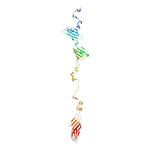
    6CL5, 6CL6

  • PubMed Abstract: 

    The R-type pyocins are high-molecular weight bacteriocins produced by some strains of Pseudomonas aeruginosa to specifically kill other strains of the same species. Structurally, the R-type pyocins are similar to "simple" contractile tails, such as those of phage P2 and Mu. The pyocin recognizes and binds to its target with the help of fibers that emanate from the baseplate structure at one end of the particle. Subsequently, the pyocin contracts its sheath and drives the rigid tube through the host cell envelope. This causes depolarization of the cytoplasmic membrane and cell death. The host cell surface-binding fiber is ~340 Å-long and is attached to the baseplate with its N-terminal domain. Here, we report the crystal structures of C-terminal fragments of the R1 and R2 pyocin fibers that comprise the distal, receptor-binding part of the protein. Both proteins are ~240 Å-long homotrimers in which slender rod-like domains are interspersed with more globular domains-two tandem knob domains in the N-terminal part of the fragment and a lectin-like domain at its C-terminus. The putative substrate binding sites are separated by about 100 Å, suggesting that binding of the fiber to the cell surface causes the fiber to adopt a certain orientation relative to the baseplate and this then triggers sheath contraction.


  • Organizational Affiliation

    Institute of Physics of Biologic Systems, École Polytechnique Fédérale de Lausanne (EPFL), BSP-415, 1015 Lausanne, Switzerland. sebuth@utmb.edu.


Macromolecules
Find similar proteins by:  (by identity cutoff)  |  3D Structure
Entity ID: 1
MoleculeChains Sequence LengthOrganismDetailsImage
Tail fiber protein
A, B, C, D, E
A, B, C, D, E, F
372Pseudomonas aeruginosa PAO1Mutation(s): 0 
Gene Names: PA0620
UniProt
Find proteins for G3XD71 (Pseudomonas aeruginosa (strain ATCC 15692 / DSM 22644 / CIP 104116 / JCM 14847 / LMG 12228 / 1C / PRS 101 / PAO1))
Explore G3XD71 
Go to UniProtKB:  G3XD71
Entity Groups  
Sequence Clusters30% Identity50% Identity70% Identity90% Identity95% Identity100% Identity
UniProt GroupG3XD71
Sequence Annotations
Expand
  • Reference Sequence
Small Molecules
Ligands 4 Unique
IDChains Name / Formula / InChI Key2D Diagram3D Interactions
EDO
Query on EDO

Download Ideal Coordinates CCD File 
AA [auth D]
CA [auth E]
DA [auth E]
EA [auth E]
FA [auth E]
AA [auth D],
CA [auth E],
DA [auth E],
EA [auth E],
FA [auth E],
HA [auth E],
IA [auth F],
J [auth A],
JA [auth F],
K [auth A],
KA [auth F],
LA [auth F],
M [auth A],
N [auth B],
O [auth B],
P [auth B],
R [auth B],
S [auth C],
T [auth C],
U [auth C],
X [auth D],
Y [auth D]
1,2-ETHANEDIOL
C2 H6 O2
LYCAIKOWRPUZTN-UHFFFAOYSA-N
FE
Query on FE

Download Ideal Coordinates CCD File 
G [auth A],
V [auth D]
FE (III) ION
Fe
VTLYFUHAOXGGBS-UHFFFAOYSA-N
CA
Query on CA

Download Ideal Coordinates CCD File 
GA [auth E]
I [auth A]
L [auth A]
Q [auth B]
W [auth D]
GA [auth E],
I [auth A],
L [auth A],
Q [auth B],
W [auth D],
Z [auth D]
CALCIUM ION
Ca
BHPQYMZQTOCNFJ-UHFFFAOYSA-N
MG
Query on MG

Download Ideal Coordinates CCD File 
BA [auth E],
H [auth A]
MAGNESIUM ION
Mg
JLVVSXFLKOJNIY-UHFFFAOYSA-N
Experimental Data & Validation

Experimental Data

  • Method: X-RAY DIFFRACTION
  • Resolution: 1.90 Å
  • R-Value Free: 0.208 
  • R-Value Work: 0.160 
  • R-Value Observed: 0.160 
  • Space Group: P 21 21 21
Unit Cell:
Length ( Å )Angle ( ˚ )
a = 56.32α = 90
b = 126.946β = 90
c = 432.938γ = 90
Software Package:
Software NamePurpose
PHENIXrefinement
XDSdata reduction
XDSdata scaling
PHASERphasing

Structure Validation

View Full Validation Report


Entry History & Funding Information

Deposition Data


Funding OrganizationLocationGrant Number
SNSFSwitzerland31003A_127092
EPFL running cost grantSwitzerland577-1

Revision History  (Full details and data files)

  • Version 1.0: 2018-06-27
    Type: Initial release
  • Version 1.1: 2019-07-17
    Changes: Data collection, Database references
  • Version 1.2: 2024-03-13
    Changes: Data collection, Database references, Derived calculations
  • Version 1.3: 2024-04-03
    Changes: Refinement description