6CH2

Crystal structure of the cytoplasmic domain of FlhA and FliT-FliD complex


Experimental Data Snapshot

  • Method: X-RAY DIFFRACTION
  • Resolution: 2.70 Å
  • R-Value Free: 0.266 
  • R-Value Work: 0.233 
  • R-Value Observed: 0.235 

wwPDB Validation   3D Report Full Report


This is version 1.2 of the entry. See complete history


Literature

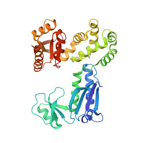
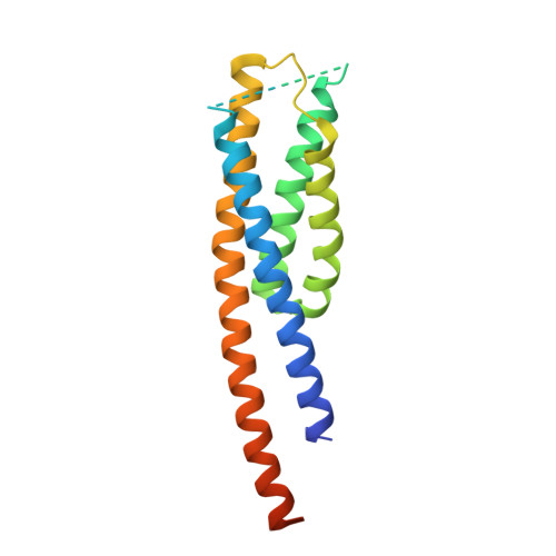
Structures of chaperone-substrate complexes docked onto the export gate in a type III secretion system.

Xing, Q.Shi, K.Portaliou, A.Rossi, P.Economou, A.Kalodimos, C.G.

(2018) Nat Commun 9: 1773-1773

  • DOI: https://doi.org/10.1038/s41467-018-04137-4
  • Primary Citation of Related Structures:  
    6CH1, 6CH2, 6CH3

  • PubMed Abstract: 

    The flagellum and the injectisome enable bacterial locomotion and pathogenesis, respectively. These nanomachines assemble and function using a type III secretion system (T3SS). Exported proteins are delivered to the export apparatus by dedicated cytoplasmic chaperones for their transport through the membrane. The structural and mechanistic basis of this process is poorly understood. Here we report the structures of two ternary complexes among flagellar chaperones (FliT and FliS), protein substrates (the filament-capping FliD and flagellin FliC), and the export gate platform protein FlhA. The substrates do not interact directly with FlhA; however, they are required to induce a binding-competent conformation to the chaperone that exposes the recognition motif featuring a highly conserved sequence recognized by FlhA. The structural data reveal the recognition signal in a class of T3SS proteins and provide new insight into the assembly of key protein complexes at the export gate.


  • Organizational Affiliation

    Department of Structural Biology, St. Jude Children's Research Hospital, 263 Danny Thomas Place, Memphis, TN, 38105, USA.


Macromolecules
Find similar proteins by:  (by identity cutoff)  |  3D Structure
Entity ID: 1
MoleculeChains Sequence LengthOrganismDetailsImage
Flagellar biosynthesis protein FlhA
A, B, C
334Salmonella enterica subsp. enterica serovar Typhimurium str. LT2Mutation(s): 0 
Gene Names: flhASTM1913
UniProt
Find proteins for P40729 (Salmonella typhimurium (strain LT2 / SGSC1412 / ATCC 700720))
Explore P40729 
Go to UniProtKB:  P40729
Entity Groups  
Sequence Clusters30% Identity50% Identity70% Identity90% Identity95% Identity100% Identity
UniProt GroupP40729
Sequence Annotations
Expand
  • Reference Sequence
Find similar proteins by:  (by identity cutoff)  |  3D Structure
Entity ID: 2
MoleculeChains Sequence LengthOrganismDetailsImage
Flagellar hook-associated protein 2,Flagellar protein FliT
D, E, F
179Salmonella enterica subsp. enterica serovar Typhimurium str. LT2Salmonella enterica subsp. enterica serovar Typhi
This entity is chimeric
Mutation(s): 0 
Gene Names: fliDflaVflbCSTM1960fliTSTY2170t0915
UniProt
Find proteins for P0A1N3 (Salmonella typhi)
Explore P0A1N3 
Go to UniProtKB:  P0A1N3
Find proteins for P16328 (Salmonella typhimurium (strain LT2 / SGSC1412 / ATCC 700720))
Explore P16328 
Go to UniProtKB:  P16328
Entity Groups  
Sequence Clusters30% Identity50% Identity70% Identity90% Identity95% Identity100% Identity
UniProt GroupsP0A1N3P16328
Sequence Annotations
Expand
  • Reference Sequence
Experimental Data & Validation

Experimental Data

  • Method: X-RAY DIFFRACTION
  • Resolution: 2.70 Å
  • R-Value Free: 0.266 
  • R-Value Work: 0.233 
  • R-Value Observed: 0.235 
  • Space Group: P 1
Unit Cell:
Length ( Å )Angle ( ˚ )
a = 49.11α = 86.88
b = 77.59β = 89
c = 119.27γ = 84.27
Software Package:
Software NamePurpose
PHENIXrefinement
XDSdata reduction
XDSdata scaling
PHASERphasing

Structure Validation

View Full Validation Report


Entry History & Funding Information

Deposition Data


Funding OrganizationLocationGrant Number
National Institutes of Health/National Institute of General Medical Sciences (NIH/NIGMS)United StatesGM118047
National Institutes of Health/National Institute of General Medical Sciences (NIH/NIGMS)United StatesGM073854

Revision History  (Full details and data files)

  • Version 1.0: 2018-05-16
    Type: Initial release
  • Version 1.1: 2020-01-01
    Changes: Author supporting evidence
  • Version 1.2: 2023-10-04
    Changes: Data collection, Database references, Refinement description