6CD1

Crystal structure of Medicago truncatula serine hydroxymethyltransferase 3 (MtSHMT3), complexes with reaction intermediates


Experimental Data Snapshot

  • Method: X-RAY DIFFRACTION
  • Resolution: 1.91 Å
  • R-Value Free: 0.236 
  • R-Value Work: 0.191 
  • R-Value Observed: 0.191 

wwPDB Validation   3D Report Full Report


Ligand Structure Quality Assessment 


This is version 1.3 of the entry. See complete history


Literature

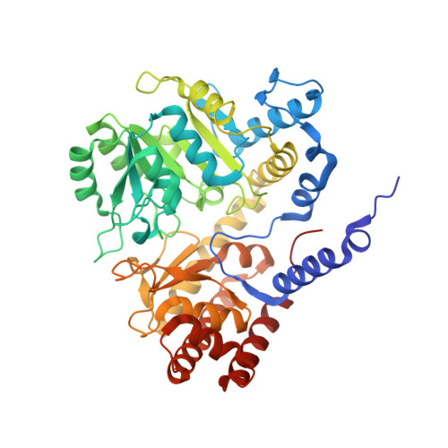
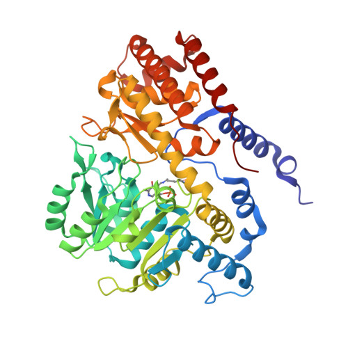
Chloroplastic Serine Hydroxymethyltransferase FromMedicago truncatula: A Structural Characterization.

Ruszkowski, M.Sekula, B.Ruszkowska, A.Dauter, Z.

(2018) Front Plant Sci 9: 584-584

  • DOI: https://doi.org/10.3389/fpls.2018.00584
  • Primary Citation of Related Structures:  
    6CCZ, 6CD0, 6CD1

  • PubMed Abstract: 

    Serine hydroxymethyltransferase (SHMT, EC 2.1.2.1) is a pyridoxal 5'-phosphate (PLP)-dependent enzyme which catalyzes the reversible serine-to-glycine conversion in either a tetrahydrofolate-dependent or -independent manner. The enzyme is also responsible for the tetrahydrofolate-independent cleavage of other β-hydroxy amino acids. In addition to being an essential player in the serine homeostasis, SHMT action is the main source of activated one-carbon units, which links SHMT activity with the control of cell proliferation. In plants, studies of SHMT enzymes are more complicated than of those of, e.g., bacterial or mammalian origins because plant genomes encode multiple SHMT isozymes that are targeted to different subcellular compartments: cytosol, mitochondria, plastids, and nucleus. Here we report crystal structures of chloroplast-targeted SHMT from Medicago truncatula ( Mt SHMT3). Mt SHMT3 is a tetramer in solution, composed of two tight and obligate dimers. Our complexes with PLP internal aldimine, PLP-serine and PLP-glycine external aldimines, and PLP internal aldimine with a free glycine reveal structural details of the Mt SHMT3-catalyzed reaction. Capturing the enzyme in different stages along the course of the slow tetrahydrofolate-independent serine-to-glycine conversion allowed to observe a unique conformation of the PLP-serine γ-hydroxyl group, and a concerted movement of two tyrosine residues in the active site.


  • Organizational Affiliation

    Synchrotron Radiation Research Section of MCL, National Cancer Institute, Argonne, IL, United States.


Macromolecules
Find similar proteins by:  (by identity cutoff)  |  3D Structure
Entity ID: 1
MoleculeChains Sequence LengthOrganismDetailsImage
Serine hydroxymethyltransferase
A, B, C, D, F
455Medicago truncatulaMutation(s): 0 
Gene Names: 11445860MTR_2g018290
EC: 2.1.2.1
UniProt
Find proteins for G7ILW0 (Medicago truncatula)
Explore G7ILW0 
Go to UniProtKB:  G7ILW0
Entity Groups  
Sequence Clusters30% Identity50% Identity70% Identity90% Identity95% Identity100% Identity
UniProt GroupG7ILW0
Sequence Annotations
Expand
  • Reference Sequence
Find similar proteins by:  (by identity cutoff)  |  3D Structure
Entity ID: 2
MoleculeChains Sequence LengthOrganismDetailsImage
Serine hydroxymethyltransferase
E, G, H
455Medicago truncatulaMutation(s): 0 
Gene Names: 11445860MTR_2g018290
EC: 2.1.2.1
UniProt
Find proteins for G7ILW0 (Medicago truncatula)
Explore G7ILW0 
Go to UniProtKB:  G7ILW0
Entity Groups  
Sequence Clusters30% Identity50% Identity70% Identity90% Identity95% Identity100% Identity
UniProt GroupG7ILW0
Sequence Annotations
Expand
  • Reference Sequence
Small Molecules
Ligands 5 Unique
IDChains Name / Formula / InChI Key2D Diagram3D Interactions
PLS (Subject of Investigation/LOI)
Query on PLS

Download Ideal Coordinates CCD File 
I [auth A],
P [auth D]
[3-HYDROXY-2-METHYL-5-PHOSPHONOOXYMETHYL-PYRIDIN-4-YLMETHYL]-SERINE
C11 H17 N2 O8 P
ODVKKQWXKRZJLG-VIFPVBQESA-N
PLG (Subject of Investigation/LOI)
Query on PLG

Download Ideal Coordinates CCD File 
K [auth B],
T [auth F]
N-GLYCINE-[3-HYDROXY-2-METHYL-5-PHOSPHONOOXYMETHYL-PYRIDIN-4-YL-METHANE]
C10 H15 N2 O7 P
FEVQWBMNLWUBTF-UHFFFAOYSA-N
GLY
Query on GLY

Download Ideal Coordinates CCD File 
R [auth E],
X [auth H]
GLYCINE
C2 H5 N O2
DHMQDGOQFOQNFH-UHFFFAOYSA-N
EDO
Query on EDO

Download Ideal Coordinates CCD File 
J [auth A]
L [auth B]
O [auth C]
Q [auth D]
S [auth E]
J [auth A],
L [auth B],
O [auth C],
Q [auth D],
S [auth E],
U [auth F],
V [auth G],
Y [auth H]
1,2-ETHANEDIOL
C2 H6 O2
LYCAIKOWRPUZTN-UHFFFAOYSA-N
ACT
Query on ACT

Download Ideal Coordinates CCD File 
M [auth C],
N [auth C],
W [auth H]
ACETATE ION
C2 H3 O2
QTBSBXVTEAMEQO-UHFFFAOYSA-M
Modified Residues  1 Unique
IDChains TypeFormula2D DiagramParent
LLP
Query on LLP
E, G, H
L-PEPTIDE LINKINGC14 H22 N3 O7 PLYS
Experimental Data & Validation

Experimental Data

  • Method: X-RAY DIFFRACTION
  • Resolution: 1.91 Å
  • R-Value Free: 0.236 
  • R-Value Work: 0.191 
  • R-Value Observed: 0.191 
  • Space Group: P 1 21 1
Unit Cell:
Length ( Å )Angle ( ˚ )
a = 94.086α = 90
b = 103.653β = 97.38
c = 180.379γ = 90
Software Package:
Software NamePurpose
REFMACrefinement
XDSdata reduction
XDSdata scaling
PHASERphasing

Structure Validation

View Full Validation Report


Ligand Structure Quality Assessment 


Entry History 

Deposition Data

Revision History  (Full details and data files)

  • Version 1.0: 2018-05-23
    Type: Initial release
  • Version 1.1: 2018-06-20
    Changes: Data collection, Database references
  • Version 1.2: 2023-10-04
    Changes: Data collection, Database references, Refinement description
  • Version 1.3: 2023-11-15
    Changes: Data collection