6CC2

Crystal Structure of CDC45 from Entamoeba histolytica


Experimental Data Snapshot

  • Method: X-RAY DIFFRACTION
  • Resolution: 1.66 Å
  • R-Value Free: 0.191 
  • R-Value Work: 0.157 
  • R-Value Observed: 0.159 

wwPDB Validation   3D Report Full Report


This is version 1.1 of the entry. See complete history


Literature

Crystal Structure ofEntamoeba histolyticaCdc45 Suggests a Conformational Switch that May Regulate DNA Replication.

Kurniawan, F.Shi, K.Kurahashi, K.Bielinsky, A.K.Aihara, H.

(2018) iScience 3: 102-109

  • DOI: https://doi.org/10.1016/j.isci.2018.04.011
  • Primary Citation of Related Structures:  
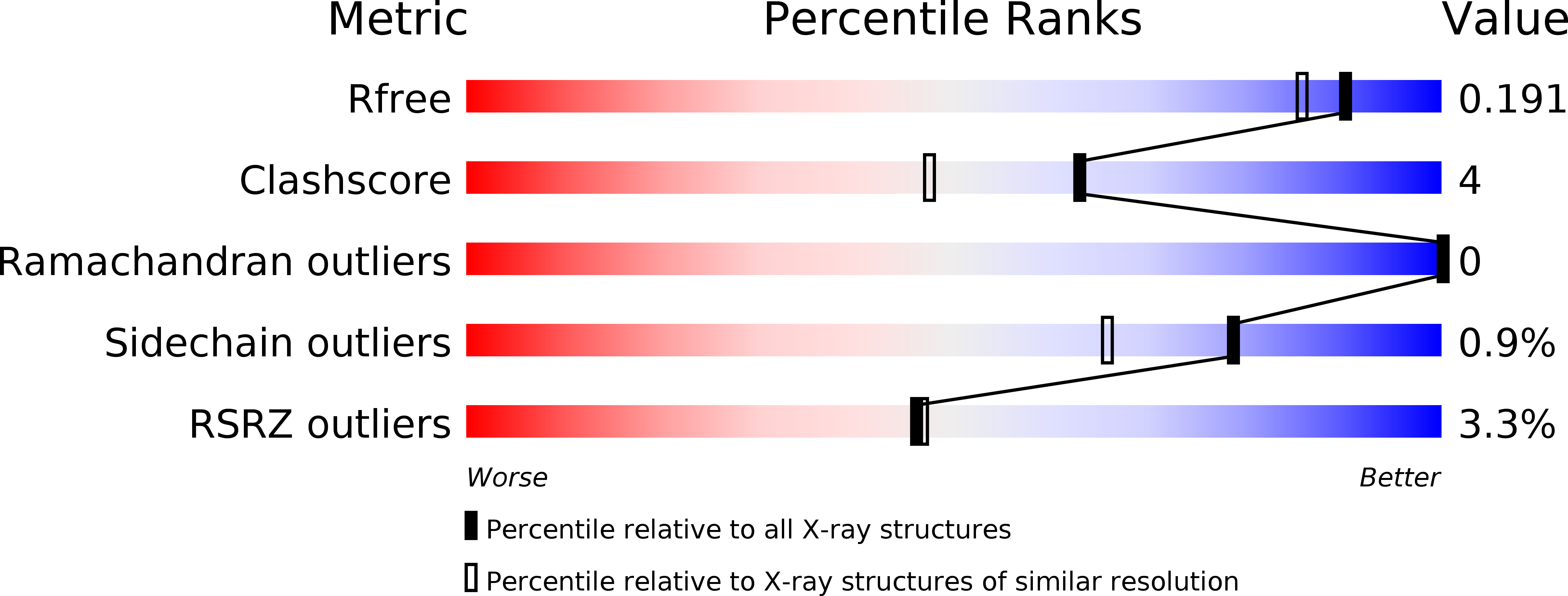
    6CC2

  • PubMed Abstract: 

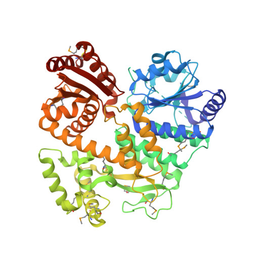
    Cdc45 plays a critical role at the core of the eukaryotic DNA replisome, serving as an essential scaffolding component of the replicative helicase holoenzyme Cdc45-MCM-GINS (CMG) complex. A 1.66-Å-resolution crystal structure of the full-length Cdc45 protein from Entamoeba histolytica shows a protein fold similar to that observed previously for human Cdc45 in its active conformation, featuring the overall disk-like monomer shape and intimate contacts between the N- and C-terminal DHH domains. However, the E. histolytica Cdc45 structure shows several unique features, including a distinct orientation of the C-terminal DHHA1 domain, concomitant disordering of the adjacent protruding α-helical segment implicated in DNA polymerase ε interactions, and a unique conformation of the GINS/Mcm5-binding loop. These structural observations collectively suggest the possibility that Cdc45 can sample multiple conformations corresponding to different functional states. We propose that such conformational switch of Cdc45 may allow regulation of protein-protein interactions important in DNA replication.


  • Organizational Affiliation

    Department of Biochemistry, Molecular Biology and Biophysics, University of Minnesota, Minneapolis, MN 55455, USA.


Macromolecules
Find similar proteins by:  (by identity cutoff)  |  3D Structure
Entity ID: 1
MoleculeChains Sequence LengthOrganismDetailsImage
Cell division control protein 45 cdc45 putative543Entamoeba histolyticaMutation(s): 0 
Gene Names: CL6EHI_049900
UniProt
Find proteins for C4LUI8 (Entamoeba histolytica (strain ATCC 30459 / HM-1:IMSS / ABRM))
Explore C4LUI8 
Go to UniProtKB:  C4LUI8
Entity Groups  
Sequence Clusters30% Identity50% Identity70% Identity90% Identity95% Identity100% Identity
UniProt GroupC4LUI8
Sequence Annotations
Expand
  • Reference Sequence
Small Molecules
Ligands 3 Unique
IDChains Name / Formula / InChI Key2D Diagram3D Interactions
PEG
Query on PEG

Download Ideal Coordinates CCD File 
Q [auth A]DI(HYDROXYETHYL)ETHER
C4 H10 O3
MTHSVFCYNBDYFN-UHFFFAOYSA-N
EDO
Query on EDO

Download Ideal Coordinates CCD File 
B [auth A]
C [auth A]
D [auth A]
E [auth A]
F [auth A]
B [auth A],
C [auth A],
D [auth A],
E [auth A],
F [auth A],
G [auth A],
H [auth A],
I [auth A],
J [auth A],
K [auth A],
L [auth A],
M [auth A]
1,2-ETHANEDIOL
C2 H6 O2
LYCAIKOWRPUZTN-UHFFFAOYSA-N
CL
Query on CL

Download Ideal Coordinates CCD File 
N [auth A],
O [auth A],
P [auth A]
CHLORIDE ION
Cl
VEXZGXHMUGYJMC-UHFFFAOYSA-M
Modified Residues  2 Unique
IDChains TypeFormula2D DiagramParent
CME
Query on CME
A
L-PEPTIDE LINKINGC5 H11 N O3 S2CYS
MSE
Query on MSE
A
L-PEPTIDE LINKINGC5 H11 N O2 SeMET
Experimental Data & Validation

Experimental Data

  • Method: X-RAY DIFFRACTION
  • Resolution: 1.66 Å
  • R-Value Free: 0.191 
  • R-Value Work: 0.157 
  • R-Value Observed: 0.159 
  • Space Group: P 21 21 21
Unit Cell:
Length ( Å )Angle ( ˚ )
a = 39.12α = 90
b = 73.81β = 90
c = 171.6γ = 90
Software Package:
Software NamePurpose
PHENIXrefinement
XDSdata reduction
XDSdata scaling
AutoSolphasing

Structure Validation

View Full Validation Report


Entry History & Funding Information

Deposition Data


Funding OrganizationLocationGrant Number
National Institutes of Health/National Institute of General Medical Sciences (NIH/NIGMS)United StatesNIGMS P41-GM103403

Revision History  (Full details and data files)

  • Version 1.0: 2018-06-27
    Type: Initial release
  • Version 1.1: 2020-01-01
    Changes: Author supporting evidence