6CBN

x-ray structure of NeoB from streptomyces fradiae in complex with PLP and neomycin (as the external aldimine) at pH 7.5


Experimental Data Snapshot

  • Method: X-RAY DIFFRACTION
  • Resolution: 1.35 Å
  • R-Value Free: 0.197 
  • R-Value Work: 0.172 
  • R-Value Observed: 0.173 

wwPDB Validation   3D Report Full Report


Ligand Structure Quality Assessment 


This is version 1.5 of the entry. See complete history


Literature

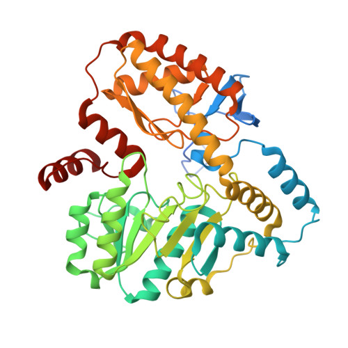
The three-dimensional structure of NeoB: An aminotransferase involved in the biosynthesis of neomycin.

Dow, G.T.Thoden, J.B.Holden, H.M.

(2018) Protein Sci 27: 945-956

  • DOI: https://doi.org/10.1002/pro.3400
  • Primary Citation of Related Structures:  
    6CBK, 6CBL, 6CBM, 6CBN, 6CBO

  • PubMed Abstract: 

    The aminoglycoside antibiotics, discovered as natural products in the 1940s, demonstrate a broad antimicrobial spectrum. Due to their nephrotoxic and ototoxic side effects, however, their widespread clinical usage has typically been limited to the treatment of serious infections. Neomycin B, first isolated from strains of Streptomyces in 1948, is one such drug that was approved for human use by the U.S. Food and Drug Administration in 1964. Only within the last 11 years has the biochemical pathway for its production been elaborated, however. Here we present the three-dimensional architecture of NeoB from Streptomyces fradiae, which is a pyridoxal 5'-phosphate or PLP-dependent aminotransferase that functions on two different substrates in neomycin B biosynthesis. For this investigation, four high resolution X-ray structures of NeoB were determined in various complexed states. The overall fold of NeoB is that typically observed for members of the "aspartate aminotransferase" family with the exception of an additional three-stranded antiparallel β-sheet that forms part of the subunit-subunit interface of the dimer. The manner in which the active site of NeoB accommodates quite different substrates has been defined by this investigation. In addition, during the course of this study, we also determined the structure of the aminotransferase GenB1 to high resolution. GenB1 functions as an aminotransferase in gentamicin biosynthesis. Taken together, the structures of NeoB and GenB1, presented here, provide the first detailed descriptions of aminotransferases that specifically function on aldehyde moieties in aminoglycoside biosynthesis.


  • Organizational Affiliation

    Department of Biochemistry, University of Wisconsin, Madison, WI, 53706.


Macromolecules
Find similar proteins by:  (by identity cutoff)  |  3D Structure
Entity ID: 1
MoleculeChains Sequence LengthOrganismDetailsImage
Neamine transaminase NeoN
A, B
438Streptomyces fradiaeMutation(s): 0 
Gene Names: neoNneo18neoB
EC: 2.6.1.93 (PDB Primary Data), 2.6.1.95 (PDB Primary Data)
UniProt
Find proteins for Q53U08 (Streptomyces fradiae)
Explore Q53U08 
Go to UniProtKB:  Q53U08
Entity Groups  
Sequence Clusters30% Identity50% Identity70% Identity90% Identity95% Identity100% Identity
UniProt GroupQ53U08
Sequence Annotations
Expand
  • Reference Sequence
Small Molecules
Ligands 2 Unique
IDChains Name / Formula / InChI Key2D Diagram3D Interactions
OZY
Query on OZY

Download Ideal Coordinates CCD File 
C [auth A],
E [auth B]
(1R,2R,3S,4R,6S)-4,6-diamino-2-[(3-O-{2-amino-2,6-dideoxy-6-[({3-hydroxy-2-methyl-5-[(phosphonooxy)methyl]pyridin-4-yl}methyl)amino]-alpha-D-glucopyranosyl}-beta-D-ribofuranosyl)oxy]-3-hydroxycyclohexyl 2,6-diamino-2,6-dideoxy-alpha-D-glucopyranoside
C31 H56 N7 O18 P
XKWYFXMYGMVOPV-AWIIFLAXSA-N
EDO
Query on EDO

Download Ideal Coordinates CCD File 
D [auth A]1,2-ETHANEDIOL
C2 H6 O2
LYCAIKOWRPUZTN-UHFFFAOYSA-N
Experimental Data & Validation

Experimental Data

  • Method: X-RAY DIFFRACTION
  • Resolution: 1.35 Å
  • R-Value Free: 0.197 
  • R-Value Work: 0.172 
  • R-Value Observed: 0.173 
  • Space Group: P 1 21 1
Unit Cell:
Length ( Å )Angle ( ˚ )
a = 59.255α = 90
b = 109.576β = 110.5
c = 70.443γ = 90
Software Package:
Software NamePurpose
REFMACrefinement
SAINTdata reduction
SADABSdata scaling
PHASERphasing

Structure Validation

View Full Validation Report


Ligand Structure Quality Assessment 


Entry History & Funding Information

Deposition Data


Funding OrganizationLocationGrant Number
National Institutes of Health/National Institute of General Medical Sciences (NIH/NIGMS)United StatesGM115921

Revision History  (Full details and data files)

  • Version 1.0: 2018-02-21
    Type: Initial release
  • Version 1.1: 2018-02-28
    Changes: Author supporting evidence
  • Version 1.2: 2018-03-21
    Changes: Database references
  • Version 1.3: 2018-05-09
    Changes: Data collection, Database references
  • Version 1.4: 2020-01-01
    Changes: Author supporting evidence
  • Version 1.5: 2023-10-04
    Changes: Data collection, Database references, Refinement description