6C9D

Crystal structure of KA1-autoinhibited MARK1 kinase

  • Classification: TRANSFERASE
  • Organism(s): Homo sapiens
  • Expression System: Escherichia coli
  • Mutation(s): Yes 

  • Deposited: 2018-01-26 Released: 2018-07-11 
  • Deposition Author(s): Emptage, R.P., Marmorstein, R.
  • Funding Organization(s): National Institutes of Health/National Institute of General Medical Sciences (NIH/NIGMS), National Institutes of Health/National Cancer Institute (NIH/NCI)

Experimental Data Snapshot

  • Method: X-RAY DIFFRACTION
  • Resolution: 2.50 Å
  • R-Value Free: 0.251 
  • R-Value Work: 0.203 
  • R-Value Observed: 0.206 

wwPDB Validation   3D Report Full Report


This is version 1.3 of the entry. See complete history


Literature

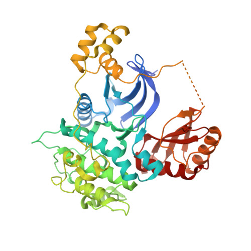
Structural Basis for MARK1 Kinase Autoinhibition by Its KA1 Domain.

Emptage, R.P.Lemmon, M.A.Ferguson, K.M.Marmorstein, R.

(2018) Structure 26: 1137

  • DOI: https://doi.org/10.1016/j.str.2018.05.008
  • Primary Citation of Related Structures:  
    6C9D

  • PubMed Abstract: 

    The kinase associated-1 (KA1) domain is found at the C-terminus of multiple Ser/Thr protein kinases from yeast to humans, and has been assigned autoinhibitory, membrane-binding, and substrate-targeting roles. Here, we report the crystal structure of the MARK1 kinase/UBA domain bound to its autoinhibitory KA1 domain, revealing an unexpected interface at the αD helix and contacts with both the N- and C-lobes of the kinase domain. We confirm the binding interface location in kinetic studies of variants mutated on the kinase domain surface. Together with other MARK kinase structures, the data implicate that the KA1 domain blocks peptide substrate binding. The structure highlights the kinase-specific autoinhibitory binding modes of different KA1 domains, and provides potential new avenues by which to intervene therapeutically in Alzheimer's disease and cancers in which MARK1 or related kinases are implicated.


  • Organizational Affiliation

    Department of Biochemistry and Biophysics and the Abramson Family Cancer Research Institute, Perelman School of Medicine, University of Pennsylvania, Philadelphia, PA 19104, USA. Electronic address: emptage@upenn.edu.


Macromolecules
Find similar proteins by:  (by identity cutoff)  |  3D Structure
Entity ID: 1
MoleculeChains Sequence LengthOrganismDetailsImage
Serine/threonine-protein kinase MARK1,Serine/threonine-protein kinase MARK1
A, B
459Homo sapiensMutation(s): 3 
Gene Names: MARK1KIAA1477MARK
EC: 2.7.11.1 (PDB Primary Data), 2.7.11.26 (PDB Primary Data)
UniProt & NIH Common Fund Data Resources
Find proteins for Q9P0L2 (Homo sapiens)
Explore Q9P0L2 
Go to UniProtKB:  Q9P0L2
PHAROS:  Q9P0L2
GTEx:  ENSG00000116141 
Entity Groups  
Sequence Clusters30% Identity50% Identity70% Identity90% Identity95% Identity100% Identity
UniProt GroupQ9P0L2
Sequence Annotations
Expand
  • Reference Sequence
Experimental Data & Validation

Experimental Data

  • Method: X-RAY DIFFRACTION
  • Resolution: 2.50 Å
  • R-Value Free: 0.251 
  • R-Value Work: 0.203 
  • R-Value Observed: 0.206 
  • Space Group: C 1 2 1
Unit Cell:
Length ( Å )Angle ( ˚ )
a = 169.993α = 90
b = 69.578β = 124.3
c = 103.999γ = 90
Software Package:
Software NamePurpose
PHENIXrefinement
PDB_EXTRACTdata extraction
HKL-2000data reduction
HKL-2000data scaling
PHASERphasing

Structure Validation

View Full Validation Report


Entry History & Funding Information

Deposition Data


Funding OrganizationLocationGrant Number
National Institutes of Health/National Institute of General Medical Sciences (NIH/NIGMS)United StatesF32GM115098
National Institutes of Health/National Cancer Institute (NIH/NCI)United States5P01CA114046

Revision History  (Full details and data files)

  • Version 1.0: 2018-07-11
    Type: Initial release
  • Version 1.1: 2018-08-29
    Changes: Data collection, Database references
  • Version 1.2: 2019-12-04
    Changes: Author supporting evidence
  • Version 1.3: 2023-10-04
    Changes: Data collection, Database references, Refinement description