6C0Y

Lysinoalanine synthase, DurN, from duramycin biosynthesis bound to duramycin


Experimental Data Snapshot

  • Method: X-RAY DIFFRACTION
  • Resolution: 1.66 Å
  • R-Value Free: 0.242 
  • R-Value Work: 0.184 
  • R-Value Observed: 0.187 

wwPDB Validation   3D Report Full Report


This is version 1.5 of the entry. See complete history


Literature

Substrate-assisted enzymatic formation of lysinoalanine in duramycin.

An, L.Cogan, D.P.Navo, C.D.Jimenez-Oses, G.Nair, S.K.van der Donk, W.A.

(2018) Nat Chem Biol 14: 928-933

  • DOI: https://doi.org/10.1038/s41589-018-0122-4
  • Primary Citation of Related Structures:  
    6C0G, 6C0H, 6C0Y

  • PubMed Abstract: 

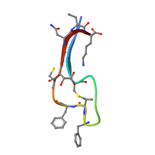
    Duramycin is a heavily post-translationally modified peptide that binds phosphatidylethanolamine. It has been investigated as an antibiotic, an inhibitor of viral entry, a therapeutic for cystic fibrosis, and a tumor and vasculature imaging agent. Duramycin contains a β-hydroxylated Asp (Hya) and four macrocycles, including an essential lysinoalanine (Lal) cross-link. The mechanism of Lal formation is not known. Here we show that Lal is installed stereospecifically by DurN via addition of Lys19 to a dehydroalanine. The structure of DurN reveals an unusual dimer with a new fold. Surprisingly, in the structure of duramycin bound to DurN, no residues of the enzyme are near the Lal cross-link. Instead, Hya15 of the substrate makes interactions with Lal, suggesting it acts as a base to deprotonate Lys19 during catalysis. Biochemical data suggest that DurN preorganizes the reactive conformation of the substrate, such that the Hya15 of the substrate can serve as the catalytic base for Lal formation.


  • Organizational Affiliation

    Department of Chemistry, University of Illinois at Urbana-Champaign, Champaign, IL, USA.


Macromolecules
Find similar proteins by:  (by identity cutoff)  |  3D Structure
Entity ID: 1
MoleculeChains Sequence LengthOrganismDetailsImage
Lysinoalanine synthase
A, B, C, D, E
A, B, C, D, E, F, G, H
121Streptomyces cinnamoneusMutation(s): 0 
Gene Names: durN
UniProt
Find proteins for A0A3F2YLX1 (Streptomyces cinnamoneus)
Explore A0A3F2YLX1 
Go to UniProtKB:  A0A3F2YLX1
Entity Groups  
Sequence Clusters30% Identity50% Identity70% Identity90% Identity95% Identity100% Identity
UniProt GroupA0A3F2YLX1
Sequence Annotations
Expand
  • Reference Sequence

Find similar proteins by:  Sequence   |   3D Structure  

Entity ID: 2
MoleculeChains Sequence LengthOrganismDetailsImage
CYS-LYS-GLN-DAL-CYS-ALA-PHE-GLY-PRO-PHE-DBB-PHE-VAL-CYS-BH2-GLY-ASN-DBB-LYS19Streptomyces cinnamoneusMutation(s): 0 
UniProt
Find proteins for A0A3F2YLX2 (Streptomyces cinnamoneus)
Explore A0A3F2YLX2 
Go to UniProtKB:  A0A3F2YLX2
Entity Groups  
Sequence Clusters30% Identity50% Identity70% Identity90% Identity95% Identity100% Identity
UniProt GroupA0A3F2YLX2
Sequence Annotations
Expand
  • Reference Sequence
Experimental Data & Validation

Experimental Data

  • Method: X-RAY DIFFRACTION
  • Resolution: 1.66 Å
  • R-Value Free: 0.242 
  • R-Value Work: 0.184 
  • R-Value Observed: 0.187 
  • Space Group: P 1
Unit Cell:
Length ( Å )Angle ( ˚ )
a = 59.881α = 71.51
b = 67.423β = 76.25
c = 69.122γ = 72.92
Software Package:
Software NamePurpose
REFMACrefinement
XDSdata reduction
XDSdata scaling
PHASERphasing

Structure Validation

View Full Validation Report


Entry History 

Deposition Data

Revision History  (Full details and data files)

  • Version 1.0: 2018-09-05
    Type: Initial release
  • Version 1.1: 2018-09-19
    Changes: Data collection, Database references
  • Version 1.2: 2018-10-03
    Changes: Data collection, Database references
  • Version 1.3: 2020-08-12
    Changes: Derived calculations, Structure summary
  • Version 1.4: 2023-10-04
    Changes: Data collection, Database references, Refinement description
  • Version 1.5: 2023-11-15
    Changes: Data collection, Derived calculations