


Crystal Structure of Glucose-6-phosphate Isomerase from Elizabethkingia anophelis
Dranow, D.M., Mayclin, S.J., Lorimer, D.D., Edwards, T.E.To be published.
Experimental Data Snapshot
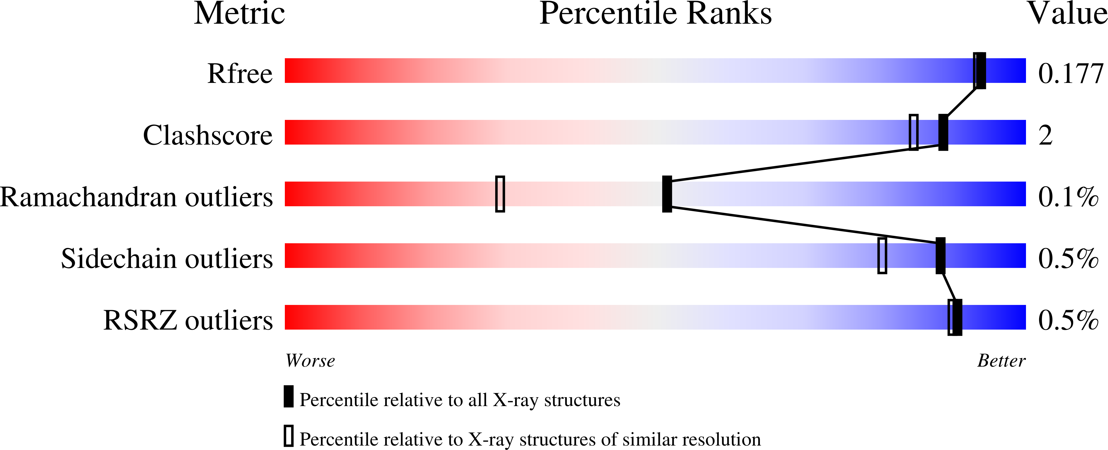
wwPDB Validation 3D Report Full Report
Entity ID: 1 | |||||
|---|---|---|---|---|---|
| Molecule | Chains | Sequence Length | Organism | Details | Image |
| Glucose-6-phosphate isomerase | 555 | Elizabethkingia anophelis NUHP1 | Mutation(s): 0 Gene Names: pgi, BD94_3890 EC: 5.3.1.9 | 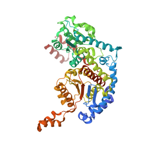 | |
UniProt | |||||
Find proteins for A0A077EQ93 (Elizabethkingia anophelis NUHP1) Explore A0A077EQ93 Go to UniProtKB: A0A077EQ93 | |||||
Entity Groups | |||||
| Sequence Clusters | 30% Identity50% Identity70% Identity90% Identity95% Identity100% Identity | ||||
| UniProt Group | A0A077EQ93 | ||||
Sequence AnnotationsExpand | |||||
| |||||
| Length ( Å ) | Angle ( ˚ ) |
|---|---|
| a = 69.95 | α = 90 |
| b = 104.75 | β = 90 |
| c = 152.65 | γ = 90 |
| Software Name | Purpose |
|---|---|
| XDS | data reduction |
| XSCALE | data scaling |
| PHENIX | refinement |
| PDB_EXTRACT | data extraction |
| MoRDa | phasing |
RCSB PDB (citation) is hosted by
RCSB PDB is a member of the

