6BWC

X-ray structure of Pen from Bacillus thuringiensis


Experimental Data Snapshot

  • Method: X-RAY DIFFRACTION
  • Resolution: 2.70 Å
  • R-Value Free: 0.251 
  • R-Value Work: 0.201 
  • R-Value Observed: 0.204 

wwPDB Validation   3D Report Full Report


Ligand Structure Quality Assessment 


This is version 1.3 of the entry. See complete history


Literature

Molecular architectures of Pen and Pal: Key enzymes required for CMP-pseudaminic acid biosynthesis in Bacillus thuringiensis.

Delvaux, N.A.Thoden, J.B.Holden, H.M.

(2018) Protein Sci 27: 738-749

  • DOI: https://doi.org/10.1002/pro.3368
  • Primary Citation of Related Structures:  
    6BWC, 6BWL

  • PubMed Abstract: 

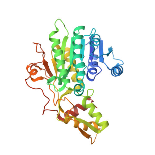
    Bacillus thuringiensis is a soil-dwelling Gram positive bacterium that has been utilized as a biopesticide for well over 60 years. It is known to contain flagella that are important for motility. One of the proteins found in flagella is flagellin, which is post-translationally modified by O-glycosylation with derivatives of pseudaminic acid. The biosynthetic pathway for the production of CMP-pseudaminic acid in B. thuringiensis, starting with UDP-N-acetyl-d-glucosamine (UDP-GlcNAc), requires seven enzymes. Here, we report the three-dimensional structures of Pen and Pal, which catalyze the first and second steps, respectively. Pen contains a tightly bound NADP(H) cofactor whereas Pal is isolated with bound NAD(H). For the X-ray analysis of Pen, the site-directed D128N/K129A mutant variant was prepared in order to trap its substrate, UDP-GlcNAc, into the active site. Pen adopts a hexameric quaternary structure with each subunit showing the bilobal architecture observed for members of the short-chain dehydrogenase/reductase superfamily. The hexameric quaternary structure is atypical for most members of the superfamily. The structure of Pal was determined in the presence of UDP. Pal adopts the more typical dimeric quaternary structure. Taken together, Pen and Pal catalyze the conversion of UDP-GlcNAc to UDP-4-keto-6-deoxy-l-N-acetylaltrosamine. Strikingly, in Gram negative bacteria such as Campylobacter jejuni and Helicobacter pylori, only a single enzyme (FlaA1) is required for the production of UDP-4-keto-6-deoxy-l-N-acetylaltrosamine. A comparison of Pen and Pal with FlaA1 reveals differences that may explain why FlaA1 is a bifunctional enzyme whereas Pen and Pal catalyze the individual steps leading to the formation of the UDP-sugar product. This investigation represents the first structural analysis of the enzymes in B. thuringiensis that are required for CMP-pseudaminic acid formation.


  • Organizational Affiliation

    Department of Biochemistry, University of Wisconsin, Madison, Wisconsin, 53706.


Macromolecules
Find similar proteins by:  (by identity cutoff)  |  3D Structure
Entity ID: 1
MoleculeChains Sequence LengthOrganismDetailsImage
Polysaccharide biosynthesis protein CapD
A, B, C, D, E
A, B, C, D, E, F
343Bacillus thuringiensis HD-771Mutation(s): 2 
Gene Names: BTG_02000
Entity Groups  
Sequence Clusters30% Identity50% Identity70% Identity90% Identity95% Identity100% Identity
Sequence Annotations
Expand
  • Reference Sequence
Small Molecules
Ligands 3 Unique
IDChains Name / Formula / InChI Key2D Diagram3D Interactions
NAP
Query on NAP

Download Ideal Coordinates CCD File 
G [auth A]
K [auth B]
N [auth C]
P [auth D]
R [auth E]
G [auth A],
K [auth B],
N [auth C],
P [auth D],
R [auth E],
T [auth F]
NADP NICOTINAMIDE-ADENINE-DINUCLEOTIDE PHOSPHATE
C21 H28 N7 O17 P3
XJLXINKUBYWONI-NNYOXOHSSA-N
UD1
Query on UD1

Download Ideal Coordinates CCD File 
H [auth A]
L [auth B]
O [auth C]
Q [auth D]
S [auth E]
H [auth A],
L [auth B],
O [auth C],
Q [auth D],
S [auth E],
U [auth F]
URIDINE-DIPHOSPHATE-N-ACETYLGLUCOSAMINE
C17 H27 N3 O17 P2
LFTYTUAZOPRMMI-CFRASDGPSA-N
SO4
Query on SO4

Download Ideal Coordinates CCD File 
I [auth A],
J [auth A],
M [auth B],
V [auth F],
W [auth F]
SULFATE ION
O4 S
QAOWNCQODCNURD-UHFFFAOYSA-L
Experimental Data & Validation

Experimental Data

  • Method: X-RAY DIFFRACTION
  • Resolution: 2.70 Å
  • R-Value Free: 0.251 
  • R-Value Work: 0.201 
  • R-Value Observed: 0.204 
  • Space Group: P 1 21 1
Unit Cell:
Length ( Å )Angle ( ˚ )
a = 92.524α = 90
b = 141.876β = 111.66
c = 93.244γ = 90
Software Package:
Software NamePurpose
REFMACrefinement
SAINTdata reduction
SADABSdata scaling
PHASERphasing

Structure Validation

View Full Validation Report


Ligand Structure Quality Assessment 


Entry History & Funding Information

Deposition Data


Funding OrganizationLocationGrant Number
National Institutes of Health/National Institute of General Medical Sciences (NIH/NIGMS)United StatesGM115921

Revision History  (Full details and data files)

  • Version 1.0: 2018-01-17
    Type: Initial release
  • Version 1.1: 2018-02-28
    Changes: Database references
  • Version 1.2: 2020-01-01
    Changes: Author supporting evidence
  • Version 1.3: 2023-10-04
    Changes: Data collection, Database references, Refinement description