6BT2

Structure of the human Nocturnin catalytic domain with bound sulfate anion


Experimental Data Snapshot

  • Method: X-RAY DIFFRACTION
  • Resolution: 2.41 Å
  • R-Value Free: 0.224 
  • R-Value Work: 0.161 
  • R-Value Observed: 0.164 

wwPDB Validation   3D Report Full Report


This is version 1.7 of the entry. See complete history


Literature

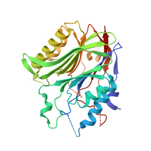
The structure of human Nocturnin reveals a conserved ribonuclease domain that represses target transcript translation and abundance in cells.

T Abshire, E.Chasseur, J.Bohn, J.A.Del Rizzo, P.A.Freddolino, P.L.Goldstrohm, A.C.Trievel, R.C.

(2018) Nucleic Acids Res 46: 6257-6270

  • DOI: https://doi.org/10.1093/nar/gky412
  • Primary Citation of Related Structures:  
    6BT1, 6BT2

  • PubMed Abstract: 

    The circadian protein Nocturnin (NOCT) belongs to the exonuclease, endonuclease and phosphatase superfamily and is most similar to the CCR4-class of deadenylases that degrade the poly-adenosine tails of mRNAs. NOCT-deficient mice are resistant to high-fat diet induced weight gain, and exhibit dysregulation of bone formation. However, the mechanisms by which NOCT regulates these processes remain to be determined. Here, we describe a pair of high-resolution crystal structures of the human NOCT catalytic domain. The active site of NOCT is highly conserved with other exoribonucleases, and when directed to a transcript in cells, NOCT can reduce translation and abundance of that mRNA in a manner dependent on key active site residues. In contrast to the related deadenylase CNOT6L, purified recombinant NOCT lacks in vitro ribonuclease activity, suggesting that unidentified factors are necessary for enzymatic activity. We also find the ability of NOCT to repress reporter mRNAs in cells depends upon the 3' end of the mRNA, as reporters terminating with a 3' MALAT1 structure cannot be repressed by NOCT. Together, these data demonstrate that NOCT is an exoribonuclease that can degrade mRNAs to inhibit protein expression, suggesting a molecular mechanism for its regulatory role in lipid metabolism and bone development.


  • Organizational Affiliation

    Department of Biological Chemistry, University of Michigan, Ann Arbor, MI 48109, USA.


Macromolecules
Find similar proteins by:  (by identity cutoff)  |  3D Structure
Entity ID: 1
MoleculeChains Sequence LengthOrganismDetailsImage
Nocturnin
A, B
313Homo sapiensMutation(s): 0 
Gene Names: NOCTCCR4CCRN4LNOC
EC: 3.1.13.4
UniProt & NIH Common Fund Data Resources
Find proteins for Q9UK39 (Homo sapiens)
Explore Q9UK39 
Go to UniProtKB:  Q9UK39
PHAROS:  Q9UK39
GTEx:  ENSG00000151014 
Entity Groups  
Sequence Clusters30% Identity50% Identity70% Identity90% Identity95% Identity100% Identity
UniProt GroupQ9UK39
Sequence Annotations
Expand
  • Reference Sequence
Experimental Data & Validation

Experimental Data

  • Method: X-RAY DIFFRACTION
  • Resolution: 2.41 Å
  • R-Value Free: 0.224 
  • R-Value Work: 0.161 
  • R-Value Observed: 0.164 
  • Space Group: P 21 21 21
Unit Cell:
Length ( Å )Angle ( ˚ )
a = 62.004α = 90
b = 69.437β = 90
c = 153.496γ = 90
Software Package:
Software NamePurpose
PHENIXrefinement
HKL-2000data collection
HKL-2000data scaling
PHENIXphasing
HKL-2000data reduction

Structure Validation

View Full Validation Report


Entry History & Funding Information

Deposition Data


Funding OrganizationLocationGrant Number
Edward Mallinckrodt Jr. FoundationUnited States--
Michigan Nutrition and Obesity Research CenterUnited States--
National Institutes of Health/National Institute of General Medical Sciences (NIH/NIGMS)United States5T32GM008597
American Heart AssociationUnited States16PRE26700002

Revision History  (Full details and data files)

  • Version 1.0: 2018-05-16
    Type: Initial release
  • Version 1.1: 2018-05-23
    Changes: Data collection, Database references
  • Version 1.2: 2018-06-20
    Changes: Data collection, Database references
  • Version 1.3: 2018-07-18
    Changes: Data collection, Database references
  • Version 1.4: 2019-02-20
    Changes: Author supporting evidence, Data collection
  • Version 1.5: 2020-01-01
    Changes: Author supporting evidence
  • Version 1.6: 2024-03-13
    Changes: Data collection, Database references, Derived calculations
  • Version 1.7: 2024-04-03
    Changes: Refinement description