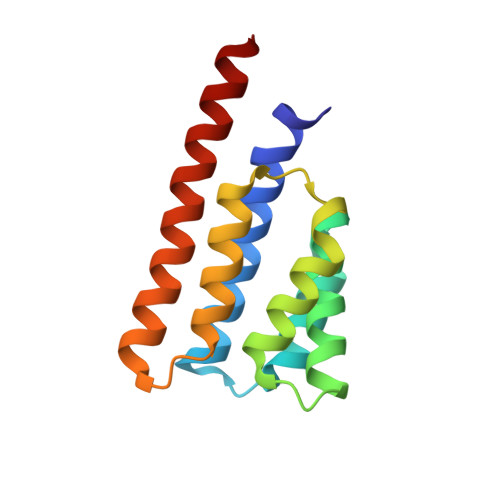
Structural characterization of SpoIIIAB sporulation-essential protein in Bacillus subtilis.
Zeytuni, N., Flanagan, K.A., Worrall, L.J., Massoni, S.C., Camp, A.H., Strynadka, N.C.J.(2018) J Struct Biol 202: 105-112
- PubMed: 29288127
- DOI: https://doi.org/10.1016/j.jsb.2017.12.009
- Primary Citation of Related Structures:
6BS9 - PubMed Abstract:
Endospore formation in the Gram-positive bacterium Bacillus subtilis initiates in response to nutrient depletion and involves a series of morphological changes that result in the creation of a dormant spore. Early in this developmental process, the cell undergoes an asymmetric cell division that produces the larger mother cell and smaller forespore, the latter destined to become the mature spore. The mother cell septal membrane then engulfs the forespore, at which time an essential channel, the so-called feeding-tube apparatus, is thought to cross both membranes to create a direct conduit between the cells. At least nine proteins are required to form this channel including SpoIIQ under forespore control and SpoIIIAA-AH under the mother cell control. Several of these proteins share similarity to components of Type-II, -III and -IV secretion systems as well as the flagellum from Gram-negative bacteria. Here we report the X-ray crystallographic structure of the cytosolic domain of SpoIIIAB to 2.3 Å resolution. This domain adopts a conserved, secretion-system related fold of a six membered anti-parallel helical bundle with a positively charged membrane-interaction face at one end and a small groove at the other end that may serve as a binding site for partner proteins in the assembled apparatus. We analyzed and identified potential interaction interfaces by structure-guided mutagenesis in vivo. Furthermore, we were able to identify a remarkable structural homology to the C-subunit of a bacterial V-ATPase. Collectively, our data provides new insight into the possible roles of SpoIIIAB protein within the secretion-like apparatus essential to bacterial sporulation.
Organizational Affiliation:
Department of Biochemistry and Molecular Biology and the Center for Blood Research, University of British Columbia, Vancouver, British Columbia, Canada.