6BS6

SusG with mixed linkage amylosaccharide


Experimental Data Snapshot

  • Method: X-RAY DIFFRACTION
  • Resolution: 2.17 Å
  • R-Value Free: 0.232 
  • R-Value Work: 0.194 
  • R-Value Observed: 0.195 

wwPDB Validation   3D Report Full Report


This is version 2.1 of the entry. See complete history


Literature

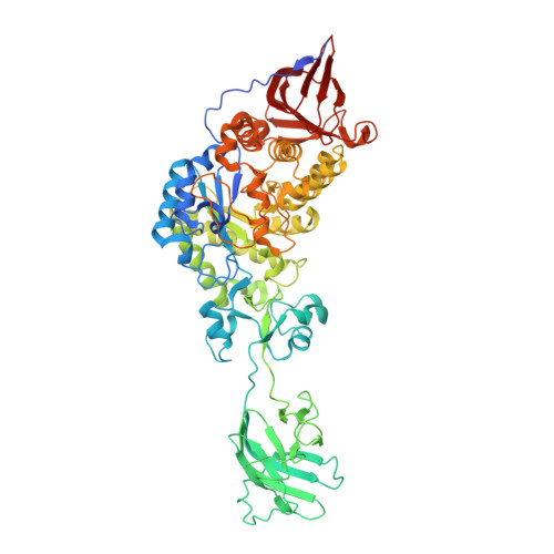
Structural basis for the flexible recognition of alpha-glucan substrates by Bacteroides thetaiotaomicron SusG.

Arnal, G.Cockburn, D.W.Brumer, H.Koropatkin, N.M.

(2018) Protein Sci 27: 1093-1101

  • DOI: https://doi.org/10.1002/pro.3410
  • Primary Citation of Related Structures:  
    6BS6

  • PubMed Abstract: 

    Bacteria that reside in the mammalian intestinal tract efficiently hydrolyze dietary carbohydrates, including starch, that escape digestion in the small intestine. Starch is an abundant dietary carbohydrate comprised of α1,4 and α1,6 linked glucose, yet mammalian intestinal glucoamylases cannot effectively hydrolyze starch that has frequent α1,6 branching as these structures hinder recognition and processing by α1,4-specific amylases. Here we present the structure of the cell surface amylase SusG from Bacteroides thetaiotaomicron complexed with a mixed linkage amylosaccharide generated from transglycosylation during crystallization. Although SusG is specific for α1,4 glucosidic bonds, binding of this new oligosaccharide at the active site demonstrates that SusG can accommodate α1,6 branch points at subsite -3 to -2, and also at subsite+1 adjacent to the site of hydrolysis, explaining how this enzyme may be able to process a wide range of limit dextrins in the intestinal environment. These data suggest that B. thetaiotaomicron and related organisms may have a selective advantage for amylosaccharide scavenging in the gut.


  • Organizational Affiliation

    Michael Smith Laboratories, University of British Columbia, 2185 East Mall, Vancouver, British Columbia, V6T 1Z4, Canada.


Macromolecules
Find similar proteins by:  (by identity cutoff)  |  3D Structure
Entity ID: 1
MoleculeChains Sequence LengthOrganismDetailsImage
Alpha-amylase SusG
A, B
670Bacteroides thetaiotaomicron VPI-5482Mutation(s): 1 
Gene Names: susGBT_3698
EC: 3.2.1.1
UniProt
Find proteins for Q8A1G3 (Bacteroides thetaiotaomicron (strain ATCC 29148 / DSM 2079 / JCM 5827 / CCUG 10774 / NCTC 10582 / VPI-5482 / E50))
Explore Q8A1G3 
Go to UniProtKB:  Q8A1G3
Entity Groups  
Sequence Clusters30% Identity50% Identity70% Identity90% Identity95% Identity100% Identity
UniProt GroupQ8A1G3
Sequence Annotations
Expand
  • Reference Sequence
Oligosaccharides

Help

Entity ID: 2
MoleculeChains Length2D Diagram Glycosylation3D Interactions
alpha-D-glucopyranose-(1-4)-alpha-D-glucopyranose-(1-4)-alpha-D-glucopyranose-(1-4)-alpha-D-glucopyranose
C, H
4N/A
Glycosylation Resources
GlyTouCan:  G87171PZ
GlyCosmos:  G87171PZ
GlyGen:  G87171PZ
Entity ID: 3
MoleculeChains Length2D Diagram Glycosylation3D Interactions
alpha-D-glucopyranose-(1-4)-alpha-D-glucopyranose-(1-6)-alpha-D-glucopyranose-(1-4)-alpha-D-glucopyranose-(1-4)-[alpha-D-glucopyranose-(1-6)]alpha-D-glucopyranose-(1-4)-alpha-D-glucopyranose-(1-4)-alpha-D-glucopyranose
D
8N/A
Glycosylation Resources
GlyTouCan:  G23144MK
GlyCosmos:  G23144MK
GlyGen:  G23144MK
Entity ID: 4
MoleculeChains Length2D Diagram Glycosylation3D Interactions
alpha-D-glucopyranose-(1-4)-[alpha-D-glucopyranose-(1-6)]alpha-D-glucopyranose-(1-4)-alpha-D-glucopyranose-(1-4)-alpha-D-glucopyranose-(1-4)-alpha-D-glucopyranose
E
6N/A
Glycosylation Resources
GlyTouCan:  G31627RU
GlyCosmos:  G31627RU
GlyGen:  G31627RU
Entity ID: 5
MoleculeChains Length2D Diagram Glycosylation3D Interactions
alpha-D-glucopyranose-(1-4)-alpha-D-glucopyranose-(1-4)-alpha-D-glucopyranose
F
3N/A
Glycosylation Resources
GlyTouCan:  G96370VA
GlyCosmos:  G96370VA
GlyGen:  G96370VA
Entity ID: 6
MoleculeChains Length2D Diagram Glycosylation3D Interactions
alpha-D-glucopyranose-(1-4)-alpha-D-glucopyranose-(1-6)-alpha-D-glucopyranose-(1-4)-alpha-D-glucopyranose-(1-4)-[alpha-D-glucopyranose-(1-6)]alpha-D-glucopyranose-(1-4)-alpha-D-glucopyranose
G
7N/A
Glycosylation Resources
GlyTouCan:  G64259JK
GlyCosmos:  G64259JK
GlyGen:  G64259JK
Experimental Data & Validation

Experimental Data

  • Method: X-RAY DIFFRACTION
  • Resolution: 2.17 Å
  • R-Value Free: 0.232 
  • R-Value Work: 0.194 
  • R-Value Observed: 0.195 
  • Space Group: P 41
Unit Cell:
Length ( Å )Angle ( ˚ )
a = 127.439α = 90
b = 127.439β = 90
c = 129.41γ = 90
Software Package:
Software NamePurpose
PHENIXrefinement
PDB_EXTRACTdata extraction
xia2data reduction
xia2data scaling
PHENIXphasing

Structure Validation

View Full Validation Report


Entry History 

Deposition Data

Revision History  (Full details and data files)

  • Version 1.0: 2018-01-24
    Type: Initial release
  • Version 1.1: 2018-04-25
    Changes: Data collection, Database references
  • Version 1.2: 2018-06-20
    Changes: Data collection, Database references
  • Version 2.0: 2020-07-29
    Type: Remediation
    Reason: Carbohydrate remediation
    Changes: Advisory, Atomic model, Data collection, Derived calculations, Structure summary
  • Version 2.1: 2023-10-04
    Changes: Data collection, Database references, Derived calculations, Refinement description, Structure summary