6BMR

Non-receptor Protein Tyrosine Phosphatase SHP2 in Complex with Allosteric Inhibitor SHP244


Experimental Data Snapshot

  • Method: X-RAY DIFFRACTION
  • Resolution: 2.21 Å
  • R-Value Free: 0.224 
  • R-Value Work: 0.167 
  • R-Value Observed: 0.170 

wwPDB Validation   3D Report Full Report


Ligand Structure Quality Assessment 


This is version 1.2 of the entry. See complete history


Literature

Dual Allosteric Inhibition of SHP2 Phosphatase.

Fodor, M.Price, E.Wang, P.Lu, H.Argintaru, A.Chen, Z.Glick, M.Hao, H.X.Kato, M.Koenig, R.LaRochelle, J.R.Liu, G.McNeill, E.Majumdar, D.Nishiguchi, G.A.Perez, L.B.Paris, G.Quinn, C.M.Ramsey, T.Sendzik, M.Shultz, M.D.Williams, S.L.Stams, T.Blacklow, S.C.Acker, M.G.LaMarche, M.J.

(2018) ACS Chem Biol 13: 647-656

  • DOI: https://doi.org/10.1021/acschembio.7b00980
  • Primary Citation of Related Structures:  
    6BMR, 6BMU, 6BMV, 6BMW, 6BMX, 6BMY

  • PubMed Abstract: 

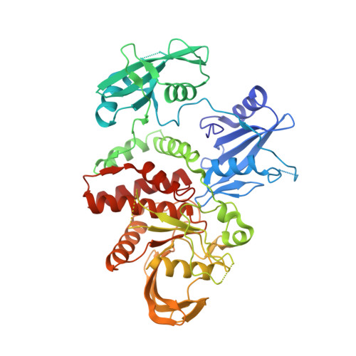
    SHP2 is a cytoplasmic protein tyrosine phosphatase encoded by the PTPN11 gene and is involved in cell proliferation, differentiation, and survival. Recently, we reported an allosteric mechanism of inhibition that stabilizes the auto-inhibited conformation of SHP2. SHP099 (1) was identified and characterized as a moderately potent, orally bioavailable, allosteric small molecule inhibitor, which binds to a tunnel-like pocket formed by the confluence of three domains of SHP2. In this report, we describe further screening strategies that enabled the identification of a second, distinct small molecule allosteric site. SHP244 (2) was identified as a weak inhibitor of SHP2 with modest thermal stabilization of the enzyme. X-ray crystallography revealed that 2 binds and stabilizes the inactive, closed conformation of SHP2, at a distinct, previously unexplored binding site-a cleft formed at the interface of the N-terminal SH2 and PTP domains. Derivatization of 2 using structure-based design resulted in an increase in SHP2 thermal stabilization, biochemical inhibition, and subsequent MAPK pathway modulation. Downregulation of DUSP6 mRNA, a downstream MAPK pathway marker, was observed in KYSE-520 cancer cells. Remarkably, simultaneous occupation of both allosteric sites by 1 and 2 was possible, as characterized by cooperative biochemical inhibition experiments and X-ray crystallography. Combining an allosteric site 1 inhibitor with an allosteric site 2 inhibitor led to enhanced pharmacological pathway inhibition in cells. This work illustrates a rare example of dual allosteric targeted protein inhibition, demonstrates screening methodology and tactics to identify allosteric inhibitors, and enables further interrogation of SHP2 in cancer and related pathologies.


  • Organizational Affiliation

    Novartis Institutes for Biomedical Research , Cambridge , Massachusetts 02139 , United States.


Macromolecules
Find similar proteins by:  (by identity cutoff)  |  3D Structure
Entity ID: 1
MoleculeChains Sequence LengthOrganismDetailsImage
Tyrosine-protein phosphatase non-receptor type 11
A, B
526Homo sapiensMutation(s): 0 
Gene Names: PTPN11PTP2CSHPTP2
EC: 3.1.3.48
UniProt & NIH Common Fund Data Resources
Find proteins for Q06124 (Homo sapiens)
Explore Q06124 
Go to UniProtKB:  Q06124
PHAROS:  Q06124
GTEx:  ENSG00000179295 
Entity Groups  
Sequence Clusters30% Identity50% Identity70% Identity90% Identity95% Identity100% Identity
UniProt GroupQ06124
Sequence Annotations
Expand
  • Reference Sequence
Small Molecules
Ligands 3 Unique
IDChains Name / Formula / InChI Key2D Diagram3D Interactions
DZV (Subject of Investigation/LOI)
Query on DZV

Download Ideal Coordinates CCD File 
C [auth A],
I [auth B]
4-[(2-chlorophenyl)methyl]-1-(2-hydroxy-3-methoxyphenyl)[1,2,4]triazolo[4,3-a]quinazolin-5(4H)-one
C23 H17 Cl N4 O3
MUHNHVCFKYIXPU-UHFFFAOYSA-N
PO4
Query on PO4

Download Ideal Coordinates CCD File 
D [auth A]
E [auth A]
F [auth A]
G [auth A]
H [auth A]
D [auth A],
E [auth A],
F [auth A],
G [auth A],
H [auth A],
J [auth B],
K [auth B],
L [auth B]
PHOSPHATE ION
O4 P
NBIIXXVUZAFLBC-UHFFFAOYSA-K
GOL
Query on GOL

Download Ideal Coordinates CCD File 
M [auth B]GLYCEROL
C3 H8 O3
PEDCQBHIVMGVHV-UHFFFAOYSA-N
Experimental Data & Validation

Experimental Data

  • Method: X-RAY DIFFRACTION
  • Resolution: 2.21 Å
  • R-Value Free: 0.224 
  • R-Value Work: 0.167 
  • R-Value Observed: 0.170 
  • Space Group: P 1 21 1
Unit Cell:
Length ( Å )Angle ( ˚ )
a = 46.23α = 90
b = 214.66β = 96.96
c = 56.03γ = 90
Software Package:
Software NamePurpose
BUSTERrefinement
XDSdata reduction
autoPROCdata scaling
BUSTERphasing

Structure Validation

View Full Validation Report


Ligand Structure Quality Assessment 


Entry History 

Deposition Data

Revision History  (Full details and data files)

  • Version 1.0: 2018-01-17
    Type: Initial release
  • Version 1.1: 2018-03-28
    Changes: Data collection, Database references
  • Version 1.2: 2023-10-04
    Changes: Data collection, Database references, Refinement description