Water-Mediated Carbon-Oxygen Hydrogen Bonding Facilitates S-Adenosylmethionine Recognition in the Reactivation Domain of Cobalamin-Dependent Methionine Synthase.
Fick, R.J., Clay, M.C., Vander Lee, L., Scheiner, S., Al-Hashimi, H., Trievel, R.C.(2018) Biochemistry 57: 3733-3740
- PubMed: 29733595
- DOI: https://doi.org/10.1021/acs.biochem.8b00375
- Primary Citation of Related Structures:
6BDY, 6BM5, 6BM6 - PubMed Abstract:
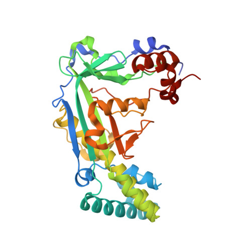
The C-terminal domain of cobalamin-dependent methionine synthase (MetH) has an essential role in catalyzing the reactivation of the enzyme following the oxidation of its cobalamin cofactor. This reactivation occurs through reductive methylation of the cobalamin using S-adenosylmethionine (AdoMet) as the methyl donor. Herein, we examine the molecular recognition of AdoMet by the MetH reactivation domain utilizing structural, biochemical, and computational approaches. Crystal structures of the Escherichia coli MetH reactivation domain in complex with AdoMet, the methyl transfer product S-adenosylhomocysteine (AdoHcy), and the AdoMet analogue inhibitor sinefungin illustrate that the ligands exhibit an analogous conformation within the solvent-exposed substrate binding cleft of the enzyme. AdoMet binding is stabilized by an intramolecular sulfur-oxygen chalcogen bond between the sulfonium and carboxylate groups of the substrate and by water-mediated carbon-oxygen hydrogen bonding between the sulfonium cation and the side chains of Glu1097 and Glu1128 that bracket the substrate binding cleft. AdoMet and sinefungin exhibited similar binding affinities for the MetH reactivation domain, whereas AdoHcy displayed an affinity for the enzyme that was an order of magnitude lower. Mutations of Glu1097 and Glu1128 diminished the AdoMet/AdoHcy binding selectivity ratio to approximately 2-fold, underscoring the role of these residues in enabling the enzyme to discriminate between the substrate and product. Together, these findings indicate that Glu1097 and Glu1128 in MetH promote high-affinity recognition of AdoMet and that sinefungin and potentially other AdoMet-based methyltransferase inhibitors can abrogate MetH reactivation, which would result in off-target effects associated with alterations in methionine homeostasis and one-carbon metabolism.
Organizational Affiliation:
Department of Biological Chemistry , University of Michigan , Ann Arbor , Michigan 48109 , United States.