6BLD

Mycobacterium marinum cytochrome P450 CYP268A2 in complex with pseudoionone


Experimental Data Snapshot

  • Method: X-RAY DIFFRACTION
  • Resolution: 2.00 Å
  • R-Value Free: 0.248 
  • R-Value Work: 0.200 
  • R-Value Observed: 0.201 

wwPDB Validation   3D Report Full Report


Ligand Structure Quality Assessment 


This is version 1.5 of the entry. See complete history


Literature

Structural and functional characterisation of the cytochrome P450 enzyme CYP268A2 from

Child, S.A.Naumann, E.F.Bruning, J.B.Bell, S.G.

(2018) Biochem J 475: 705-722

  • DOI: https://doi.org/10.1042/BCJ20170946
  • Primary Citation of Related Structures:  
    6BLD

  • PubMed Abstract: 

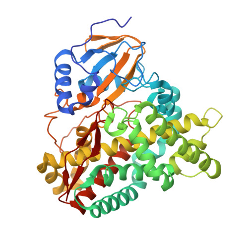
    Members of the cytochrome P450 monooxygenase family CYP268 are found across a broad range of Mycobacterium species including the pathogens Mycobacterium avium , M. colombiense , M. kansasii , and M marinum CYP268A2, from M. marinum , which is the first member of this family to be studied, was purified and characterised. CYP268A2 was found to bind a variety of substrates with high affinity, including branched and straight chain fatty acids (C10-C12), acetate esters, and aromatic compounds. The enzyme was also found to bind phenylimidazole inhibitors but not larger azoles, such as ketoconazole. The monooxygenase activity of CYP268A2 was efficiently reconstituted using heterologous electron transfer partner proteins. CYP268A2 hydroxylated geranyl acetate and tran s-pseudoionone at a terminal methyl group to yield ( 2E , 6E )-8-hydroxy-3,7-dimethylocta-2,6-dien-1-yl acetate and ( 3E , 5E , 9E )-11-hydroxy-6,10-dimethylundeca-3,5,9-trien-2-one, respectively. The X-ray crystal structure of CYP268A2 was solved to a resolution of 2.0 Å with trans -pseudoionone bound in the active site. The overall structure was similar to that of the related phytanic acid monooxygenase CYP124A1 enzyme from Mycobacterium tuberculosis , which shares 41% sequence identity. The active site is predominantly hydrophobic, but includes the Ser99 and Gln209 residues which form hydrogen bonds with the terminal carbonyl group of the pseudoionone. The structure provided an explanation on why CYP268A2 shows a preference for shorter substrates over the longer chain fatty acids which bind to CYP124A1 and the selective nature of the catalysed monooxygenase activity.


  • Organizational Affiliation

    Department of Chemistry, School of Physical Sciences, University of Adelaide, Adelaide, SA 5005, Australia.


Macromolecules
Find similar proteins by:  (by identity cutoff)  |  3D Structure
Entity ID: 1
MoleculeChains Sequence LengthOrganismDetailsImage
Cytochrome P450 268A2 Cyp268A2419Mycobacterium marinum MMutation(s): 0 
Gene Names: cyp268A2MMAR_3761
UniProt
Find proteins for B2HMF7 (Mycobacterium marinum (strain ATCC BAA-535 / M))
Explore B2HMF7 
Go to UniProtKB:  B2HMF7
Entity Groups  
Sequence Clusters30% Identity50% Identity70% Identity90% Identity95% Identity100% Identity
UniProt GroupB2HMF7
Sequence Annotations
Expand
  • Reference Sequence
Small Molecules
Ligands 2 Unique
IDChains Name / Formula / InChI Key2D Diagram3D Interactions
HEM
Query on HEM

Download Ideal Coordinates CCD File 
B [auth A]PROTOPORPHYRIN IX CONTAINING FE
C34 H32 Fe N4 O4
KABFMIBPWCXCRK-RGGAHWMASA-L
DXJ
Query on DXJ

Download Ideal Coordinates CCD File 
C [auth A](3E,5E)-6,10-dimethylundeca-3,5,9-trien-2-one
C13 H20 O
JXJIQCXXJGRKRJ-KOOBJXAQSA-N
Binding Affinity Annotations 
IDSourceBinding Affinity
DXJ Binding MOAD:  6BLD Kd: 3600 (nM) from 1 assay(s)
Experimental Data & Validation

Experimental Data

  • Method: X-RAY DIFFRACTION
  • Resolution: 2.00 Å
  • R-Value Free: 0.248 
  • R-Value Work: 0.200 
  • R-Value Observed: 0.201 
  • Space Group: C 1 2 1
Unit Cell:
Length ( Å )Angle ( ˚ )
a = 154.759α = 90
b = 44.726β = 100.84
c = 57.727γ = 90
Software Package:
Software NamePurpose
PHENIXrefinement
MOSFLMdata collection
Aimlessdata scaling
CNSphasing
REFMACphasing
Cootmodel building
PDB_EXTRACTdata extraction
XDSdata reduction

Structure Validation

View Full Validation Report


Ligand Structure Quality Assessment 


Entry History & Funding Information

Deposition Data


Funding OrganizationLocationGrant Number
Australian Research Council (ARC)AustraliaFT140100355

Revision History  (Full details and data files)

  • Version 1.0: 2018-01-24
    Type: Initial release
  • Version 1.1: 2018-01-31
    Changes: Database references
  • Version 1.2: 2018-02-21
    Changes: Database references
  • Version 1.3: 2018-02-28
    Changes: Database references
  • Version 1.4: 2020-01-01
    Changes: Author supporting evidence
  • Version 1.5: 2023-10-04
    Changes: Data collection, Database references, Refinement description