6BL7

S. cerevisiae stu2 coiled coil domain


Experimental Data Snapshot

  • Method: X-RAY DIFFRACTION
  • Resolution: 2.50 Å
  • R-Value Free: 0.292 
  • R-Value Work: 0.257 
  • R-Value Observed: 0.260 

wwPDB Validation   3D Report Full Report


This is version 1.3 of the entry. See complete history


Literature

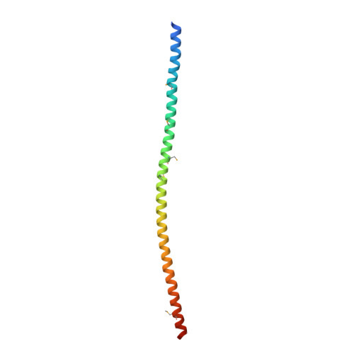
Stu2 uses a 15-nm parallel coiled coil for kinetochore localization and concomitant regulation of the mitotic spindle.

Haase, K.P.Fox, J.C.Byrnes, A.E.Adikes, R.C.Speed, S.K.Haase, J.Friedman, B.Cook, D.M.Bloom, K.Rusan, N.M.Slep, K.C.

(2018) Mol Biol Cell 29: 285-294

  • DOI: https://doi.org/10.1091/mbc.E17-01-0057
  • Primary Citation of Related Structures:  
    6BL7

  • PubMed Abstract: 

    XMAP215/Dis1 family proteins are potent microtubule polymerases, critical for mitotic spindle structure and dynamics. While microtubule polymerase activity is driven by an N-terminal tumor overexpressed gene (TOG) domain array, proper cellular localization is a requisite for full activity and is mediated by a C-terminal domain. Structural insight into the C-terminal domain's architecture and localization mechanism remain outstanding. We present the crystal structure of the Saccharomyces cerevisiae Stu2 C-terminal domain, revealing a 15-nm parallel homodimeric coiled coil. The parallel architecture of the coiled coil has mechanistic implications for the arrangement of the homodimer's N-terminal TOG domains during microtubule polymerization. The coiled coil has two spatially distinct conserved regions: CRI and CRII. Mutations in CRI and CRII perturb the distribution and localization of Stu2 along the mitotic spindle and yield defects in spindle morphology including increased frequencies of mispositioned and fragmented spindles. Collectively, these data highlight roles for the Stu2 dimerization domain as a scaffold for factor binding that optimally positions Stu2 on the mitotic spindle to promote proper spindle structure and dynamics.


  • Organizational Affiliation

    Molecular and Cellular Biophysics Program, University of North Carolina, Chapel Hill, NC 27599.


Macromolecules
Find similar proteins by:  (by identity cutoff)  |  3D Structure
Entity ID: 1
MoleculeChains Sequence LengthOrganismDetailsImage
Protein STU2
A, B
110Saccharomyces cerevisiaeMutation(s): 0 
Gene Names: STU2YLR045CL2108
UniProt
Find proteins for P46675 (Saccharomyces cerevisiae (strain ATCC 204508 / S288c))
Explore P46675 
Go to UniProtKB:  P46675
Entity Groups  
Sequence Clusters30% Identity50% Identity70% Identity90% Identity95% Identity100% Identity
UniProt GroupP46675
Sequence Annotations
Expand
  • Reference Sequence
Small Molecules
Modified Residues  1 Unique
IDChains TypeFormula2D DiagramParent
MSE
Query on MSE
A, B
L-PEPTIDE LINKINGC5 H11 N O2 SeMET
Experimental Data & Validation

Experimental Data

  • Method: X-RAY DIFFRACTION
  • Resolution: 2.50 Å
  • R-Value Free: 0.292 
  • R-Value Work: 0.257 
  • R-Value Observed: 0.260 
  • Space Group: P 1 21 1
Unit Cell:
Length ( Å )Angle ( ˚ )
a = 57.252α = 90
b = 30.112β = 102.08
c = 72.856γ = 90
Software Package:
Software NamePurpose
PHENIXrefinement
HKL-2000data collection
HKL-2000data scaling
PDB_EXTRACTdata extraction
HKL-2000data reduction
PHASERphasing

Structure Validation

View Full Validation Report


Entry History & Funding Information

Deposition Data

  • Released Date: 2017-12-13 
  • Deposition Author(s): Slep, K.C.

Funding OrganizationLocationGrant Number
National Institutes of Health/National Institute of General Medical Sciences (NIH/NIGMS)United StatesR01GM094415
National Institutes of Health/National Institute of General Medical Sciences (NIH/NIGMS)United StatesR37GM32238
National Institutes of Health/National Institute of General Medical Sciences (NIH/NIGMS)United StatesT32GM008570

Revision History  (Full details and data files)

  • Version 1.0: 2017-12-13
    Type: Initial release
  • Version 1.1: 2018-01-17
    Changes: Author supporting evidence
  • Version 1.2: 2018-02-14
    Changes: Database references
  • Version 1.3: 2020-01-01
    Changes: Author supporting evidence