6BKA

Crystal Structure of Nitronate Monooxygenase from Cyberlindnera saturnus


Experimental Data Snapshot

  • Method: X-RAY DIFFRACTION
  • Resolution: 1.65 Å
  • R-Value Free: 0.179 
  • R-Value Work: 0.140 
  • R-Value Observed: 0.142 

wwPDB Validation   3D Report Full Report


Ligand Structure Quality Assessment 


This is version 1.3 of the entry. See complete history


Literature

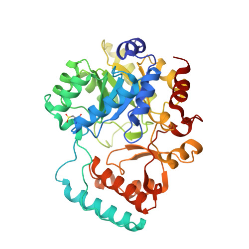
Crystal structure of yeast nitronate monooxygenase from Cyberlindnera saturnus.

Agniswamy, J.Reis, R.A.G.Wang, Y.F.Smitherman, C.Su, D.Weber, I.Gadda, G.

(2018) Proteins 86: 599-605

  • DOI: https://doi.org/10.1002/prot.25470
  • Primary Citation of Related Structures:  
    6BKA

  • PubMed Abstract: 

    Nitronate monooxygenase (NMO) is an FMN-dependent enzyme that oxidizes the neurotoxin propionate 3-nitronate (P3N) and represents the best-known system for P3N detoxification in different organisms. The crystal structure of the first eukaryotic Class I NMO from Cyberlindnera saturnus (CsNMO) has been solved at 1.65 Å resolution and refined to an R-factor of 14.0%. The three-dimensional structures of yeast CsNMO and bacterial PaNMO are highly conserved with the exception of three additional loops on the surface in the CsNMO enzyme and differences in four active sites residues. A PEG molecule was identified in the structure and formed extensive interactions with CsNMO, suggesting a specific binding site; however, 8% PEG showed no significant effect on the enzyme activity. This new crystal structure of a eukaryotic NMO provides insight into the function of this class of enzymes.


  • Organizational Affiliation

    Department of Biology, Georgia State University, Atlanta, Georgia, 30302-3965.


Macromolecules
Find similar proteins by:  (by identity cutoff)  |  3D Structure
Entity ID: 1
MoleculeChains Sequence LengthOrganismDetailsImage
Nitronate monooxygenase374Cyberlindnera mrakiiMutation(s): 0 
EC: 1.13.12.16
UniProt
Find proteins for Q12723 (Cyberlindnera mrakii)
Explore Q12723 
Go to UniProtKB:  Q12723
Entity Groups  
Sequence Clusters30% Identity50% Identity70% Identity90% Identity95% Identity100% Identity
UniProt GroupQ12723
Sequence Annotations
Expand
  • Reference Sequence
Small Molecules
Ligands 3 Unique
IDChains Name / Formula / InChI Key2D Diagram3D Interactions
33O
Query on 33O

Download Ideal Coordinates CCD File 
C [auth A]3,6,9,12,15,18,21,24,27,30,33,36-dodecaoxaoctatriacontane-1,38-diol
C26 H54 O14
AKWFJQNBHYVIPY-UHFFFAOYSA-N
FMN
Query on FMN

Download Ideal Coordinates CCD File 
B [auth A]FLAVIN MONONUCLEOTIDE
C17 H21 N4 O9 P
FVTCRASFADXXNN-SCRDCRAPSA-N
GOL
Query on GOL

Download Ideal Coordinates CCD File 
D [auth A]GLYCEROL
C3 H8 O3
PEDCQBHIVMGVHV-UHFFFAOYSA-N
Modified Residues  1 Unique
IDChains TypeFormula2D DiagramParent
CSD
Query on CSD
A
L-PEPTIDE LINKINGC3 H7 N O4 SCYS
Experimental Data & Validation

Experimental Data

  • Method: X-RAY DIFFRACTION
  • Resolution: 1.65 Å
  • R-Value Free: 0.179 
  • R-Value Work: 0.140 
  • R-Value Observed: 0.142 
  • Space Group: P 1 21 1
Unit Cell:
Length ( Å )Angle ( ˚ )
a = 44.82α = 90
b = 68.43β = 106.32
c = 58.366γ = 90
Software Package:
Software NamePurpose
REFMACrefinement
HKL-2000data reduction
SCALEPACKdata scaling
PHASERphasing

Structure Validation

View Full Validation Report


Ligand Structure Quality Assessment 


Entry History & Funding Information

Deposition Data


Funding OrganizationLocationGrant Number
National Science Foundation (NSF, United States)United StatesCHE-1506518

Revision History  (Full details and data files)

  • Version 1.0: 2018-02-14
    Type: Initial release
  • Version 1.1: 2018-05-02
    Changes: Data collection, Database references
  • Version 1.2: 2019-11-27
    Changes: Author supporting evidence
  • Version 1.3: 2023-10-04
    Changes: Data collection, Database references, Refinement description