6B16

P21-activated kinase 1 in complex with a 4-azaindole inhibitor


Experimental Data Snapshot

  • Method: X-RAY DIFFRACTION
  • Resolution: 2.29 Å
  • R-Value Free: 0.250 
  • R-Value Work: 0.202 
  • R-Value Observed: 0.204 

wwPDB Validation   3D Report Full Report


Ligand Structure Quality Assessment 


This is version 1.0 of the entry. See complete history


Literature

Synthesis and evaluation of a series of 4-azaindole-containing p21-activated kinase-1 inhibitors.

Lee, W.Crawford, J.J.Aliagas, I.Murray, L.J.Tay, S.Wang, W.Heise, C.E.Hoeflich, K.P.La, H.Mathieu, S.Mintzer, R.Ramaswamy, S.Rouge, L.Rudolph, J.

(2016) Bioorg Med Chem Lett 26: 3518-3524

  • DOI: https://doi.org/10.1016/j.bmcl.2016.06.031
  • Primary Citation of Related Structures:  
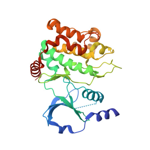
    6B16

  • PubMed Abstract: 

    A series of 4-azaindole-containing p21-activated kinase-1 (PAK1) inhibitors was prepared with the goal of improving physicochemical properties relative to an indole starting point. Indole 1 represented an attractive, non-basic scaffold with good PAK1 affinity and cellular potency but was compromised by high lipophilicity (clogD=4.4). Azaindole 5 was designed as an indole surrogate with the goal of lowering logD and resulted in equipotent PAK1 inhibition with a 2-fold improvement in cellular potency over 1. Structure-activity relationship studies around 5 identified additional 4-azaindole analogs with superior PAK1 biochemical activity (Ki <10nM) and up to 24-fold selectivity for group I over group II PAKs. Compounds from this series showed enhanced permeability, improved aqueous solubility, and lower plasma protein binding over indole 1. The improvement in physicochemical properties translated to a 20-fold decrease in unbound clearance in mouse PK studies for azaindole 5 relative to indole 1.


  • Organizational Affiliation

    Discovery Chemistry, Genentech, Inc., 1 DNA Way, South San Francisco, CA 94080, USA. Electronic address: wendyle@gene.com.


Macromolecules
Find similar proteins by:  (by identity cutoff)  |  3D Structure
Entity ID: 1
MoleculeChains Sequence LengthOrganismDetailsImage
Serine/threonine-protein kinase PAK 1
A, B
301Homo sapiensMutation(s): 0 
Gene Names: PAK1
EC: 2.7.11.1
UniProt & NIH Common Fund Data Resources
Find proteins for Q13153 (Homo sapiens)
Explore Q13153 
Go to UniProtKB:  Q13153
PHAROS:  Q13153
GTEx:  ENSG00000149269 
Entity Groups  
Sequence Clusters30% Identity50% Identity70% Identity90% Identity95% Identity100% Identity
UniProt GroupQ13153
Sequence Annotations
Expand
  • Reference Sequence
Small Molecules
Ligands 2 Unique
IDChains Name / Formula / InChI Key2D Diagram3D Interactions
C7Y
Query on C7Y

Download Ideal Coordinates CCD File 
C [auth A],
E [auth B]
N~4~-(5-cyclopropyl-1H-pyrazol-3-yl)-N~2~-[(1S)-1-(1H-pyrrolo[3,2-b]pyridin-5-yl)ethyl]pyrimidine-2,4-diamine
C19 H20 N8
GUERACYCGKAFOU-NSHDSACASA-N
SO4
Query on SO4

Download Ideal Coordinates CCD File 
D [auth A]SULFATE ION
O4 S
QAOWNCQODCNURD-UHFFFAOYSA-L
Binding Affinity Annotations 
IDSourceBinding Affinity
C7Y BindingDB:  6B16 Ki: 32 (nM) from 1 assay(s)
Binding MOAD:  6B16 Ki: 32 (nM) from 1 assay(s)
Experimental Data & Validation

Experimental Data

  • Method: X-RAY DIFFRACTION
  • Resolution: 2.29 Å
  • R-Value Free: 0.250 
  • R-Value Work: 0.202 
  • R-Value Observed: 0.204 
  • Space Group: P 1 21 1
Unit Cell:
Length ( Å )Angle ( ˚ )
a = 62.682α = 90
b = 82.458β = 106.31
c = 66.036γ = 90
Software Package:
Software NamePurpose
PHENIXrefinement
HKL-2000data reduction
SCALEPACKdata scaling
PHASERphasing

Structure Validation

View Full Validation Report


Ligand Structure Quality Assessment 


Entry History 

Deposition Data

Revision History  (Full details and data files)

  • Version 1.0: 2017-10-25
    Type: Initial release