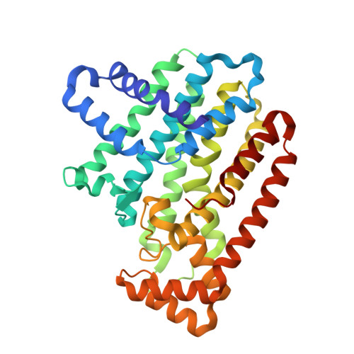
6B07

Crystal structure of CfFPPS2, a lepidopteran type-II farnesyl diphosphate synthase, complexed with [1-phosphono-2-(1-propylpyridin-2-yl)ethyl]phosphonic acid (inhibitor 1d)


Experimental Data Snapshot

  • Method: X-RAY DIFFRACTION
  • Resolution: 1.98 Å
  • R-Value Free: 0.209 
  • R-Value Work: 0.171 
  • R-Value Observed: 0.173 

wwPDB Validation   3D Report Full Report


Ligand Structure Quality Assessment 


This is version 1.4 of the entry. See complete history


Literature

Structural characterization of a lepidopteran type-II farnesyl diphosphate synthase from the spruce budworm, Choristoneura fumiferana: Implications for inhibitor design.

Picard, M.E.Nisole, A.Beliveau, C.Sen, S.Barbar, A.Shi, R.Cusson, M.

(2017) Insect Biochem Mol Biol 92: 84-92

  • DOI: https://doi.org/10.1016/j.ibmb.2017.11.011
  • Primary Citation of Related Structures:  
    6B02, 6B04, 6B06, 6B07

  • PubMed Abstract: 

    Farnesyl diphosphate synthase (FPPS) is an enzyme from the class of short chain (E)-prenyltransferases that catalyzes the condensation of two molecules of isopentenyl diphosphate (IPP, C 5 ) with dimethylallyl diphosphate (DMAPP, C 5 ) to generate the C 15 product FPP. In insects, FPPS plays a key role in the biosynthesis of the morphogenetic and gonadotropic "juvenile hormone" (JH). Lepidopteran genomes encode two very distinct FPPS paralogs, one of which ("type-II") is expressed almost exclusively in the JH-producing glands, the corpora allata. This paralog has been hypothesized to display structural features that enable the binding of the bulkier precursors required for the biosynthesis of lepidopteran ethyl-branched JHs. Here, we report on the first crystal structures of an insect FPPS solved to date. Apo, ligand-bound, and inhibitor-bound structures of type-II FPPS (FPPS2) from the spruce budworm, Choristoneura fumiferana (Order: Lepidoptera), were obtained. Comparison of apo and inhibitor-bound enzymes revealed differences in both inhibitor binding and structural plasticity of CfFPPS2 compared to other FPPSs. Our data showed that IPP is not essential to the closure of the C-terminal tail. Ortho-substituted pyridinium bisphosphonates, previously shown to inhibit CfFPPS2, bound to the allylic site, as predicted; however, their alkyl groups were oriented towards the homoallylic binding site, with the bulkier propyl-substituted inhibitor penetrating deeply into the IPP binding pocket. The current study sheds light on the structural basis of substrate specificity of type-II FPPS of the spruce budworm. Through a comparison with other inhibitor-bound FPPSs, we propose several approaches to improve inhibitor selectivity and potency.


  • Organizational Affiliation

    Département de biochimie, de microbiologie et de bio-informatique, Institut de Biologie Intégrative et des Systèmes, PROTEO, Université Laval, Quebec City, QC, G1V 0A6, Canada. Electronic address: marie-eve.picard.8@ulaval.ca.


Macromolecules
Find similar proteins by:  (by identity cutoff)  |  3D Structure
Entity ID: 1
MoleculeChains Sequence LengthOrganismDetailsImage
Farnesyl diphosphate synthase
A, B, C
341Choristoneura fumiferanaMutation(s): 0 
EC: 2.5.1
UniProt
Find proteins for Q1XAB1 (Choristoneura fumiferana)
Explore Q1XAB1 
Go to UniProtKB:  Q1XAB1
Entity Groups  
Sequence Clusters30% Identity50% Identity70% Identity90% Identity95% Identity100% Identity
UniProt GroupQ1XAB1
Sequence Annotations
Expand
  • Reference Sequence
Small Molecules
Ligands 3 Unique
IDChains Name / Formula / InChI Key2D Diagram3D Interactions
C6M
Query on C6M

Download Ideal Coordinates CCD File 
D [auth A],
J [auth B],
P [auth C]
[1-phosphono-2-(1-propylpyridin-2-yl)ethyl]phosphonic acid
C10 H18 N O6 P2
AQESRNWSIMYUEN-UHFFFAOYSA-N
EDO
Query on EDO

Download Ideal Coordinates CCD File 
H [auth A]
I [auth A]
N [auth B]
O [auth B]
T [auth C]
H [auth A],
I [auth A],
N [auth B],
O [auth B],
T [auth C],
U [auth C],
V [auth C]
1,2-ETHANEDIOL
C2 H6 O2
LYCAIKOWRPUZTN-UHFFFAOYSA-N
MG
Query on MG

Download Ideal Coordinates CCD File 
E [auth A]
F [auth A]
G [auth A]
K [auth B]
L [auth B]
E [auth A],
F [auth A],
G [auth A],
K [auth B],
L [auth B],
M [auth B],
Q [auth C],
R [auth C],
S [auth C]
MAGNESIUM ION
Mg
JLVVSXFLKOJNIY-UHFFFAOYSA-N
Experimental Data & Validation

Experimental Data

  • Method: X-RAY DIFFRACTION
  • Resolution: 1.98 Å
  • R-Value Free: 0.209 
  • R-Value Work: 0.171 
  • R-Value Observed: 0.173 
  • Space Group: C 1 2 1
Unit Cell:
Length ( Å )Angle ( ˚ )
a = 186.913α = 90
b = 122.579β = 106.51
c = 69.064γ = 90
Software Package:
Software NamePurpose
REFMACrefinement
SCALEPACKdata scaling
PDB_EXTRACTdata extraction
DENZOdata reduction
MOLREPphasing

Structure Validation

View Full Validation Report


Ligand Structure Quality Assessment 


Entry History & Funding Information

Deposition Data


Funding OrganizationLocationGrant Number
Natural Sciences and Engineering Research Council (NSERC, Canada)Canada436202

Revision History  (Full details and data files)

  • Version 1.0: 2017-12-13
    Type: Initial release
  • Version 1.1: 2018-01-10
    Changes: Database references
  • Version 1.2: 2018-01-17
    Changes: Author supporting evidence
  • Version 1.3: 2020-01-08
    Changes: Author supporting evidence
  • Version 1.4: 2023-10-04
    Changes: Data collection, Database references, Derived calculations, Refinement description