6AZT

Asparaginyl endopeptidase 1 bound to AAN peptide, a tetrahedral intermediate


Experimental Data Snapshot

  • Method: X-RAY DIFFRACTION
  • Resolution: 1.80 Å
  • R-Value Free: 0.189 
  • R-Value Work: 0.152 
  • R-Value Observed: 0.154 

wwPDB Validation   3D Report Full Report


This is version 3.0 of the entry. See complete history


Literature

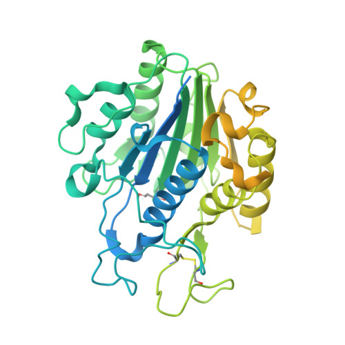
Structural basis of ribosomal peptide macrocyclization in plants.

Haywood, J.Schmidberger, J.W.James, A.M.Nonis, S.G.Sukhoverkov, K.V.Elias, M.Bond, C.S.Mylne, J.S.

(2018) Elife 7

  • DOI: https://doi.org/10.7554/eLife.32955
  • Primary Citation of Related Structures:  
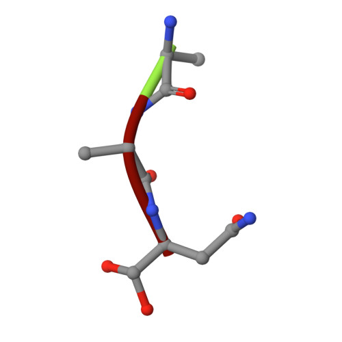
    6AZT

  • PubMed Abstract: 

    Constrained, cyclic peptides encoded by plant genes represent a new generation of drug leads. Evolution has repeatedly recruited the Cys-protease asparaginyl endopeptidase (AEP) to perform their head-to-tail ligation. These macrocyclization reactions use the substrates amino terminus instead of water to deacylate, so a peptide bond is formed. How solvent-exposed plant AEPs macrocyclize is poorly understood. Here we present the crystal structure of an active plant AEP from the common sunflower, Helianthus annuus . The active site contained electron density for a tetrahedral intermediate with partial occupancy that predicted a binding mode for peptide macrocyclization. By substituting catalytic residues we could alter the ratio of cyclic to acyclic products. Moreover, we showed AEPs from other species lacking cyclic peptides can perform macrocyclization under favorable pH conditions. This structural characterization of AEP presents a logical framework for engineering superior enzymes that generate macrocyclic peptide drug leads.


  • Organizational Affiliation

    School of Molecular Sciences, The University of Western Australia, Perth, Australia.


Macromolecules
Find similar proteins by:  (by identity cutoff)  |  3D Structure
Entity ID: 1
MoleculeChains Sequence LengthOrganismDetailsImage
Asparaginyl endopeptidase 1491Helianthus annuusMutation(s): 0 
Gene Names: AEP1
UniProt
Find proteins for A0A251RPF5 (Helianthus annuus)
Explore A0A251RPF5 
Go to UniProtKB:  A0A251RPF5
Entity Groups  
Sequence Clusters30% Identity50% Identity70% Identity90% Identity95% Identity100% Identity
UniProt GroupA0A251RPF5
Sequence Annotations
Expand
  • Reference Sequence

Find similar proteins by:  Sequence   |   3D Structure  

Entity ID: 2
MoleculeChains Sequence LengthOrganismDetailsImage
ALA-ALA-ASN tetrahedral intermediate3synthetic constructMutation(s): 0 
Sequence Annotations
Expand
  • Reference Sequence
Small Molecules
Modified Residues  2 Unique
IDChains TypeFormula2D DiagramParent
SNN
Query on SNN
A
L-PEPTIDE LINKINGC4 H6 N2 O2ASN
CE7
Query on CE7
B
L-PEPTIDE LINKINGC4 H10 N2 O4ASN
Experimental Data & Validation

Experimental Data

  • Method: X-RAY DIFFRACTION
  • Resolution: 1.80 Å
  • R-Value Free: 0.189 
  • R-Value Work: 0.152 
  • R-Value Observed: 0.154 
  • Space Group: P 31 2 1
Unit Cell:
Length ( Å )Angle ( ˚ )
a = 77.03α = 90
b = 77.03β = 90
c = 108.171γ = 120
Software Package:
Software NamePurpose
REFMACrefinement
iMOSFLMdata reduction
Aimlessdata scaling
PHASERphasing

Structure Validation

View Full Validation Report


Entry History & Funding Information

Deposition Data

  • Released Date: 2018-02-07 
  • Deposition Author(s): Bond, C.S.

Funding OrganizationLocationGrant Number
Australian Research Council (ARC)AustraliaDP160100107

Revision History  (Full details and data files)

  • Version 1.0: 2018-02-07
    Type: Initial release
  • Version 2.0: 2018-05-16
    Changes: Atomic model, Data collection, Database references, Derived calculations, Non-polymer description, Polymer sequence, Structure summary
  • Version 2.1: 2020-01-01
    Changes: Author supporting evidence
  • Version 3.0: 2023-11-15
    Changes: Atomic model, Data collection, Database references, Derived calculations