6AZL

Structure of cetuximab with aminoheptanoic acid-linked N-carboxyethylarginine meditope variant


Experimental Data Snapshot

  • Method: X-RAY DIFFRACTION
  • Resolution: 2.48 Å
  • R-Value Free: 0.216 
  • R-Value Work: 0.170 
  • R-Value Observed: 0.172 

wwPDB Validation   3D Report Full Report


This is version 1.2 of the entry. See complete history


Literature

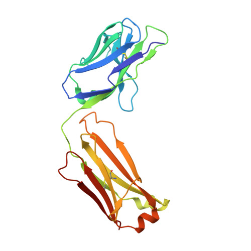
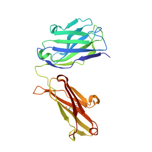
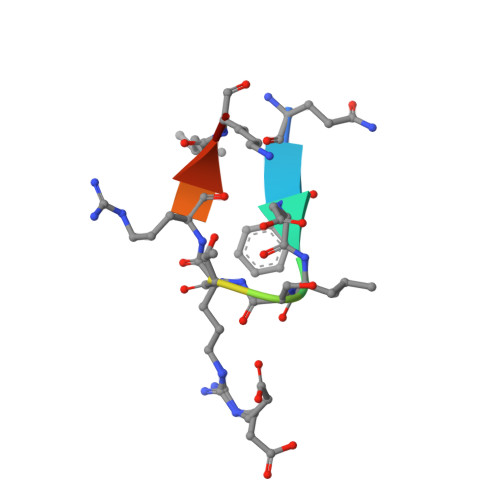
Meditope-Fab interaction: threading the hole.

Bzymek, K.P.Ma, Y.Avery, K.N.Horne, D.A.Williams, J.C.

(2017) Acta Crystallogr F Struct Biol Commun 73: 688-694

  • DOI: https://doi.org/10.1107/S2053230X17016272
  • Primary Citation of Related Structures:  
    6AU5, 6AXP, 6AYN, 6AZK, 6AZL

  • PubMed Abstract: 

    Meditope, a cyclic 12-residue peptide, binds to a unique binding side between the light and heavy chains of the cetuximab Fab. In an effort to improve the affinity of the interaction, it was sought to extend the side chain of Arg8 in the meditope, a residue that is accessible from the other side of the meditope binding site, in order to increase the number of interactions. These modifications included an n-butyl and n-octyl extension as well as hydroxyl, amine and carboxyl substitutions. The atomic structures of the complexes and the binding kinetics for each modified meditope indicated that each extension threaded through the Fab `hole' and that the carboxyethylarginine substitution makes a favorable interaction with the Fab, increasing the half-life of the complex by threefold compared with the unmodified meditope. Taken together, these studies provide a basis for the design of additional modifications to enhance the overall affinity of this unique interaction.


  • Organizational Affiliation

    Department of Molecular Medicine, Beckman Research Institute of City of Hope, 1710 Flower Street, Duarte, CA 91101, USA.


Macromolecules
Find similar proteins by:  (by identity cutoff)  |  3D Structure
Entity ID: 1
MoleculeChains Sequence LengthOrganismDetailsImage
cetuximab Fab light chain
A, C
213Mus musculusHomo sapiens
This entity is chimeric
Mutation(s): 0 
UniProt & NIH Common Fund Data Resources
Find proteins for P01834 (Homo sapiens)
Explore P01834 
Go to UniProtKB:  P01834
PHAROS:  P01834
Entity Groups  
Sequence Clusters30% Identity50% Identity70% Identity90% Identity95% Identity100% Identity
UniProt GroupP01834
Sequence Annotations
Expand
  • Reference Sequence
Find similar proteins by:  (by identity cutoff)  |  3D Structure
Entity ID: 2
MoleculeChains Sequence LengthOrganismDetailsImage
cetuximab Fab heavy chain
B, D
221Mus musculusHomo sapiens
This entity is chimeric
Mutation(s): 0 
UniProt
Find proteins for S6B291 (Homo sapiens)
Explore S6B291 
Go to UniProtKB:  S6B291
Entity Groups  
Sequence Clusters30% Identity50% Identity70% Identity90% Identity95% Identity100% Identity
UniProt GroupS6B291
Sequence Annotations
Expand
  • Reference Sequence

Find similar proteins by:  Sequence   |   3D Structure  

Entity ID: 3
MoleculeChains Sequence LengthOrganismDetailsImage
meditope
E, F
11synthetic constructMutation(s): 0 
Entity Groups  
Sequence Clusters30% Identity50% Identity70% Identity90% Identity95% Identity100% Identity
Sequence Annotations
Expand
  • Reference Sequence
Experimental Data & Validation

Experimental Data

  • Method: X-RAY DIFFRACTION
  • Resolution: 2.48 Å
  • R-Value Free: 0.216 
  • R-Value Work: 0.170 
  • R-Value Observed: 0.172 
  • Space Group: P 21 21 21
Unit Cell:
Length ( Å )Angle ( ˚ )
a = 64.57α = 90
b = 83.05β = 90
c = 212.11γ = 90
Software Package:
Software NamePurpose
PHENIXrefinement
XDSdata reduction
XSCALEdata scaling
MOLREPphasing

Structure Validation

View Full Validation Report


Entry History 

Deposition Data

Revision History  (Full details and data files)

  • Version 1.0: 2017-12-13
    Type: Initial release
  • Version 1.1: 2023-10-04
    Changes: Data collection, Database references, Refinement description
  • Version 1.2: 2023-11-15
    Changes: Data collection, Derived calculations