Structural basis for the regulation of beta-glucuronidase expression by human gut Enterobacteriaceae.
Little, M.S., Pellock, S.J., Walton, W.G., Tripathy, A., Redinbo, M.R.(2018) Proc Natl Acad Sci U S A 115: E152-E161
- PubMed: 29269393
- DOI: https://doi.org/10.1073/pnas.1716241115
- Primary Citation of Related Structures:
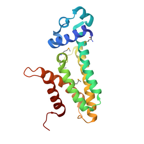
6AYH, 6AYI, 6AZ6, 6AZH - PubMed Abstract:
The gut microbiota harbor diverse β-glucuronidase (GUS) enzymes that liberate glucuronic acid (GlcA) sugars from small-molecule conjugates and complex carbohydrates. However, only the Enterobacteriaceae family of human gut-associated Proteobacteria maintain a GUS operon under the transcriptional control of a glucuronide repressor, GusR. Despite its potential importance in Escherichia , Salmonella , Klebsiella , Shigella , and Yersinia opportunistic pathogens, the structure of GusR has not been examined. Here, we explore the molecular basis for GusR-mediated regulation of GUS expression in response to small-molecule glucuronides. Presented are 2.1-Å-resolution crystal structures of GusRs from Escherichia coli and Salmonella enterica in complexes with a glucuronide ligand. The GusR-specific DNA operator site in the regulatory region of the E. coli GUS operon is identified, and structure-guided GusR mutants pinpoint the residues essential for DNA binding and glucuronide recognition. Interestingly, the endobiotic estradiol-17-glucuronide and the xenobiotic indomethacin-acyl-glucuronide are found to exhibit markedly differential binding to these GusR orthologs. Using structure-guided mutations, we are able to transfer E. coli GusR's preferential DNA and glucuronide binding affinity to S. enterica GusR. Structures of putative GusR orthologs from GUS-encoding Firmicutes species also reveal functionally unique features of the Enterobacteriaceae GusRs. Finally, dominant-negative GusR variants are validated in cell-based studies. These data provide a molecular framework toward understanding the control of glucuronide utilization by opportunistic pathogens in the human gut.
Organizational Affiliation:
Department of Chemistry, University of North Carolina at Chapel Hill, Chapel Hill, NC 27599-3290.