6AWP

X-ray structure of the ts3 human serotonin transporter complexed with fluvoxamine at the central site


Experimental Data Snapshot

  • Method: X-RAY DIFFRACTION
  • Resolution: 3.80 Å
  • R-Value Free: 0.269 
  • R-Value Work: 0.245 
  • R-Value Observed: 0.247 

wwPDB Validation   3D Report Full Report


Ligand Structure Quality Assessment 


This is version 1.7 of the entry. See complete history


Literature

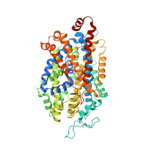
Structural basis for recognition of diverse antidepressants by the human serotonin transporter.

Coleman, J.A.Gouaux, E.

(2018) Nat Struct Mol Biol 25: 170-175

  • DOI: https://doi.org/10.1038/s41594-018-0026-8
  • Primary Citation of Related Structures:  
    6AWN, 6AWO, 6AWP, 6AWQ

  • PubMed Abstract: 

    Selective serotonin reuptake inhibitors are clinically prescribed antidepressants that act by increasing the local concentrations of neurotransmitters at synapses and in extracellular spaces via blockade of the serotonin transporter. Here we report X-ray structures of engineered thermostable variants of the human serotonin transporter bound to the antidepressants sertraline, fluvoxamine, and paroxetine. The drugs prevent serotonin binding by occupying the central substrate-binding site and stabilizing the transporter in an outward-open conformation. These structures explain how residues within the central site orchestrate binding of chemically diverse inhibitors and mediate transporter drug selectivity.


  • Organizational Affiliation

    Vollum Institute, Oregon Health & Science University, Portland, OR, USA.


Macromolecules
Find similar proteins by:  (by identity cutoff)  |  3D Structure
Entity ID: 1
MoleculeChains Sequence LengthOrganismDetailsImage
Sodium-dependent serotonin transporter549Homo sapiensMutation(s): 5 
Gene Names: SLC6A4HTTSERT
Membrane Entity: Yes 
UniProt & NIH Common Fund Data Resources
Find proteins for P31645 (Homo sapiens)
Explore P31645 
Go to UniProtKB:  P31645
PHAROS:  P31645
GTEx:  ENSG00000108576 
Entity Groups  
Sequence Clusters30% Identity50% Identity70% Identity90% Identity95% Identity100% Identity
UniProt GroupP31645
Sequence Annotations
Expand
  • Reference Sequence
Find similar proteins by:  (by identity cutoff)  |  3D Structure
Entity ID: 2
MoleculeChains Sequence LengthOrganismDetailsImage
8B6 antibody FAB heavy chain229Mus musculusMutation(s): 0 
Membrane Entity: Yes 
UniProt
Find proteins for A0A0F7R1P3 (Mus musculus)
Explore A0A0F7R1P3 
Go to UniProtKB:  A0A0F7R1P3
Entity Groups  
Sequence Clusters30% Identity50% Identity70% Identity90% Identity95% Identity100% Identity
UniProt GroupA0A0F7R1P3
Sequence Annotations
Expand
  • Reference Sequence
Find similar proteins by:  (by identity cutoff)  |  3D Structure
Entity ID: 3
MoleculeChains Sequence LengthOrganismDetailsImage
8B6 antibody FAB light chain214Mus musculusMutation(s): 0 
Membrane Entity: Yes 
UniProt
Find proteins for A0A0F7R5U8 (Mus musculus)
Explore A0A0F7R5U8 
Go to UniProtKB:  A0A0F7R5U8
Entity Groups  
Sequence Clusters30% Identity50% Identity70% Identity90% Identity95% Identity100% Identity
UniProt GroupA0A0F7R5U8
Sequence Annotations
Expand
  • Reference Sequence
Small Molecules
Ligands 5 Unique
IDChains Name / Formula / InChI Key2D Diagram3D Interactions
CLR
Query on CLR

Download Ideal Coordinates CCD File 
F [auth A]CHOLESTEROL
C27 H46 O
HVYWMOMLDIMFJA-DPAQBDIFSA-N
FVX
Query on FVX

Download Ideal Coordinates CCD File 
D [auth A]Fluvoxamine
C15 H21 F3 N2 O2
CJOFXWAVKWHTFT-XSFVSMFZSA-N
NAG
Query on NAG

Download Ideal Coordinates CCD File 
E [auth A],
G [auth A]
2-acetamido-2-deoxy-beta-D-glucopyranose
C8 H15 N O6
OVRNDRQMDRJTHS-FMDGEEDCSA-N
CL
Query on CL

Download Ideal Coordinates CCD File 
H [auth A]CHLORIDE ION
Cl
VEXZGXHMUGYJMC-UHFFFAOYSA-M
NA
Query on NA

Download Ideal Coordinates CCD File 
I [auth A],
J [auth A]
SODIUM ION
Na
FKNQFGJONOIPTF-UHFFFAOYSA-N
Binding Affinity Annotations 
IDSourceBinding Affinity
FVX BindingDB:  6AWP Ki: min: 1.46, max: 540 (nM) from 14 assay(s)
IC50: min: 3.8, max: 540 (nM) from 2 assay(s)
Experimental Data & Validation

Experimental Data

  • Method: X-RAY DIFFRACTION
  • Resolution: 3.80 Å
  • R-Value Free: 0.269 
  • R-Value Work: 0.245 
  • R-Value Observed: 0.247 
  • Space Group: C 2 2 21
Unit Cell:
Length ( Å )Angle ( ˚ )
a = 129.49α = 90
b = 163.86β = 90
c = 140.95γ = 90
Software Package:
Software NamePurpose
PHENIXrefinement
XDSdata reduction
XSCALEdata scaling
PHASERphasing

Structure Validation

View Full Validation Report


Ligand Structure Quality Assessment 


Entry History & Funding Information

Deposition Data


Funding OrganizationLocationGrant Number
National Institutes of Health/National Institute of Mental Health (NIH/NIMH)United States5R37MH070039

Revision History  (Full details and data files)

  • Version 1.0: 2017-10-25
    Type: Initial release
  • Version 1.1: 2017-12-06
    Changes: Author supporting evidence
  • Version 1.2: 2018-01-31
    Changes: Database references
  • Version 1.3: 2018-02-14
    Changes: Database references
  • Version 1.4: 2018-02-21
    Changes: Database references
  • Version 1.5: 2019-11-27
    Changes: Author supporting evidence
  • Version 1.6: 2020-07-29
    Type: Remediation
    Reason: Carbohydrate remediation
    Changes: Data collection, Derived calculations, Structure summary
  • Version 1.7: 2023-10-04
    Changes: Data collection, Database references, Refinement description, Structure summary