6AP1

Vps4p-Vta1p complex with peptide binding to the central pore of Vps4p


Experimental Data Snapshot

  • Method: ELECTRON MICROSCOPY
  • Resolution: 3.20 Å
  • Aggregation State: PARTICLE 
  • Reconstruction Method: SINGLE PARTICLE 

wwPDB Validation   3D Report Full Report


This is version 1.1 of the entry. See complete history


Literature

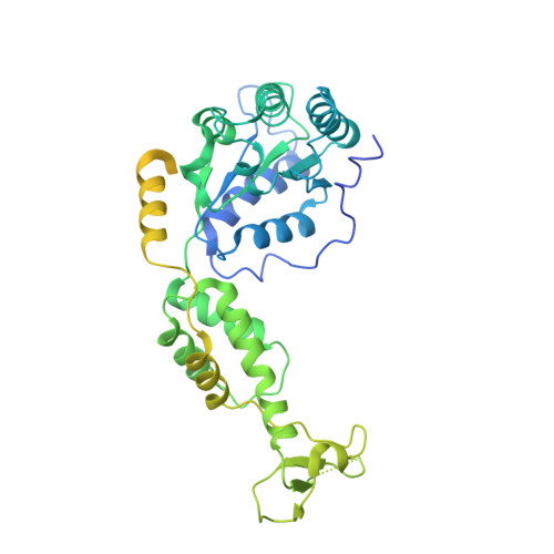
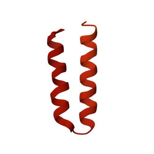
The AAA ATPase Vps4 binds ESCRT-III substrates through a repeating array of dipeptide-binding pockets.

Han, H.Monroe, N.Sundquist, W.I.Shen, P.S.Hill, C.P.

(2017) Elife 6

  • DOI: https://doi.org/10.7554/eLife.31324
  • Primary Citation of Related Structures:  
    6AP1, 6BMF

  • PubMed Abstract: 

    The hexameric AAA ATPase Vps4 drives membrane fission by remodeling and disassembling ESCRT-III filaments. Building upon our earlier 4.3 Å resolution cryo-EM structure (Monroe et al., 2017), we now report a 3.2 Å structure of Vps4 bound to an ESCRT-III peptide substrate. The new structure reveals that the peptide approximates a β-strand conformation whose helical symmetry matches that of the five Vps4 subunits it contacts directly. Adjacent Vps4 subunits make equivalent interactions with successive substrate dipeptides through two distinct classes of side chain binding pockets formed primarily by Vps4 pore loop 1. These pockets accommodate a wide range of residues, while main chain hydrogen bonds may help dictate substrate-binding orientation. The structure supports a 'conveyor belt' model of translocation in which ATP binding allows a Vps4 subunit to join the growing end of the helix and engage the substrate, while hydrolysis and release promotes helix disassembly and substrate release at the lagging end.


  • Organizational Affiliation

    Department of Biochemistry, University of Utah School of Medicine, Salt Lake City, United States.


Macromolecules
Find similar proteins by:  (by identity cutoff)  |  3D Structure
Entity ID: 1
MoleculeChains Sequence LengthOrganismDetailsImage
Vacuolar protein sorting-associated protein 4,Protein hcp1
A,
B,
C,
D,
E,
G [auth F]
519Saccharomyces cerevisiae S288CPseudomonas aeruginosa PAO1
This entity is chimeric
Mutation(s): 0 
Gene Names: 
UniProt
Find proteins for P52917 (Saccharomyces cerevisiae (strain ATCC 204508 / S288c))
Explore P52917 
Go to UniProtKB:  P52917
Find proteins for Q9I747 (Pseudomonas aeruginosa (strain ATCC 15692 / DSM 22644 / CIP 104116 / JCM 14847 / LMG 12228 / 1C / PRS 101 / PAO1))
Explore Q9I747 
Go to UniProtKB:  Q9I747
Entity Groups  
Sequence Clusters30% Identity50% Identity70% Identity90% Identity95% Identity100% Identity
UniProt GroupsP52917Q9I747
Sequence Annotations
Expand
  • Reference Sequence

Find similar proteins by:  Sequence   |   3D Structure  

Entity ID: 2
MoleculeChains Sequence LengthOrganismDetailsImage
ACE-ASP-GLU-ILE-VAL-ASN-LYS-VAL-LEU-NH2F [auth G]10Saccharomyces cerevisiaeMutation(s): 0 
UniProt
Find proteins for P36108 (Saccharomyces cerevisiae (strain ATCC 204508 / S288c))
Explore P36108 
Go to UniProtKB:  P36108
Entity Groups  
Sequence Clusters30% Identity50% Identity70% Identity90% Identity95% Identity100% Identity
UniProt GroupP36108
Sequence Annotations
Expand
  • Reference Sequence
Find similar proteins by:  (by identity cutoff)  |  3D Structure
Entity ID: 3
MoleculeChains Sequence LengthOrganismDetailsImage
Vacuolar protein sorting-associated protein VTA1
H, I, J, K, L
H, I, J, K, L, M, N, O, P, Q, R, S
330Saccharomyces cerevisiae S288CMutation(s): 0 
Gene Names: VTA1YLR181C
UniProt
Find proteins for Q06263 (Saccharomyces cerevisiae (strain ATCC 204508 / S288c))
Explore Q06263 
Go to UniProtKB:  Q06263
Entity Groups  
Sequence Clusters30% Identity50% Identity70% Identity90% Identity95% Identity100% Identity
UniProt GroupQ06263
Sequence Annotations
Expand
  • Reference Sequence
Small Molecules
Ligands 3 Unique
IDChains Name / Formula / InChI Key2D Diagram3D Interactions
ADP
Query on ADP

Download Ideal Coordinates CCD File 
CA [auth D],
EA [auth E],
T [auth A],
W [auth B],
Z [auth C]
ADENOSINE-5'-DIPHOSPHATE
C10 H15 N5 O10 P2
XTWYTFMLZFPYCI-KQYNXXCUSA-N
BEF
Query on BEF

Download Ideal Coordinates CCD File 
AA [auth C],
U [auth A],
X [auth B]
BERYLLIUM TRIFLUORIDE ION
Be F3
OGIAHMCCNXDTIE-UHFFFAOYSA-K
MG
Query on MG

Download Ideal Coordinates CCD File 
BA [auth C],
DA [auth D],
V [auth A],
Y [auth B]
MAGNESIUM ION
Mg
JLVVSXFLKOJNIY-UHFFFAOYSA-N
Experimental Data & Validation

Experimental Data

  • Method: ELECTRON MICROSCOPY
  • Resolution: 3.20 Å
  • Aggregation State: PARTICLE 
  • Reconstruction Method: SINGLE PARTICLE 
EM Software:
TaskSoftware PackageVersion
RECONSTRUCTIONRELION1.4

Structure Validation

View Full Validation Report


Entry History 

Deposition Data

Revision History  (Full details and data files)

  • Version 1.0: 2017-12-06
    Type: Initial release
  • Version 1.1: 2018-07-18
    Changes: Data collection, Experimental preparation