Structure of a fructose-1,6-bis(phosphate) aldolase liganded to its natural substrate in a cleavage-defective mutant at 2.3 A(,).
Choi, K.H., Mazurkie, A.S., Morris, A.J., Utheza, D., Tolan, D.R., Allen, K.N.(1999) Biochemistry 38: 12655-12664
- PubMed: 10504235
- DOI: https://doi.org/10.1021/bi9828371
- Primary Citation of Related Structures:
6ALD - PubMed Abstract:
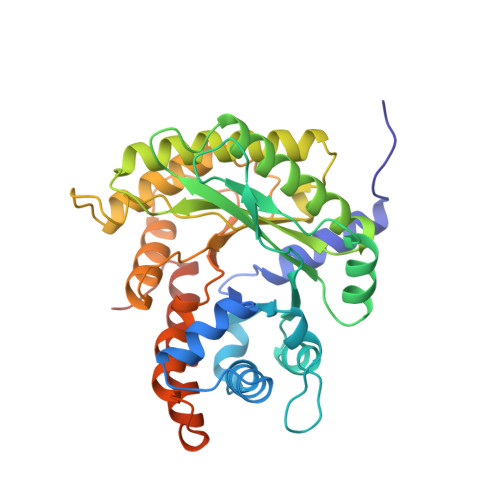
Class I fructose-1,6-bis(phosphate) aldolase is a glycolytic enzyme that catalyzes the cleavage of fructose 1,6-bis(phosphate) through a covalent Schiff base intermediate. Although the atomic structure of this enzyme is known, assigning catalytic roles to the various enzymic active-site residues has been hampered by the lack of a structure for the enzyme-substrate complex. A mutant aldolase, K146A, is unable to cleave the C3-C4 bond of the hexose while retaining the ability to form the covalent intermediate, although at a greatly diminished rate. The structure of rabbit muscle K146A-aldolase A, in complex with its native substrate, fructose 1,6-bis(phosphate), is determined to 2.3 A resolution by molecular replacement. The density at the hexose binding site differs between subunits of the tetramer, in that two sites show greater occupancy relative to the other two. The hexose is bound in its linear, open conformation, but not covalently linked to the Schiff base-forming Lys-229. Therefore, this structure most likely represents the bound complex of hexose just after hemiketal hydrolysis and prior to Schiff base formation. The C1-phosphate binding site involves the three backbone nitrogens of Ser-271, Gly-272, and Gly-302, and the epsilon-amino group of Lys-229. This is the same binding site previously found for the analogous phosphate of the product DHAP. The C6-phosphate binding site involves three basic side chains, Arg-303, Arg-42, and Lys-41. The residues closest to Lys-229 were relatively unchanged in position when compared to the unbound wild-type structure. The major differences between the bound and unbound enzyme structures were observed in the positions of Lys-107, Arg-303, and Arg-42, with the greatest difference in the change in conformation of Arg-303. Site-directed mutagenesis was performed on those residues with different conformations in bound versus unbound enzyme. The kinetic constants of these mutant enzymes with the substrates fructose 1, 6-bis(phosphate) and fructose 1-phosphate are consistent with their ligand interactions as revealed by the structure reported here, including differing effects on k(cat) and K(m) between the two substrates depending on whether the mutations affect C6-phosphate binding. In the unbound state, Arg-303 forms a salt bridge with Glu-34, and in the liganded structure it interacts closely with the substrate C6-phosphate. The position of the sugar in the binding site would require a large movement prior to achieving the proper position for covalent catalysis with the Schiff base-forming Lys-229. The movement most likely involves a change in the location of the more loosely bound C6-phosphate. This result suggests that the substrate has one position in the Michaelis complex and another in the covalent complex. Such movement could trigger conformational changes in the carboxyl-terminal region, which has been implicated in substrate specificity.
Organizational Affiliation:
Structural Biology Group, Department of Physiology, Boston University School of Medicine, 80 East Concord Street, Boston, Massachusetts 02118-2394, USA.