


Structural basis for nucleotide-mediated remodelling mechanism of Mycobacterium Mfd
Putta, S., Prabha, S., Bhat, V., Fox, G.C., Walsh, M.A., Rao, D.N., Nagaraja, V., Natesh, R.To be published.
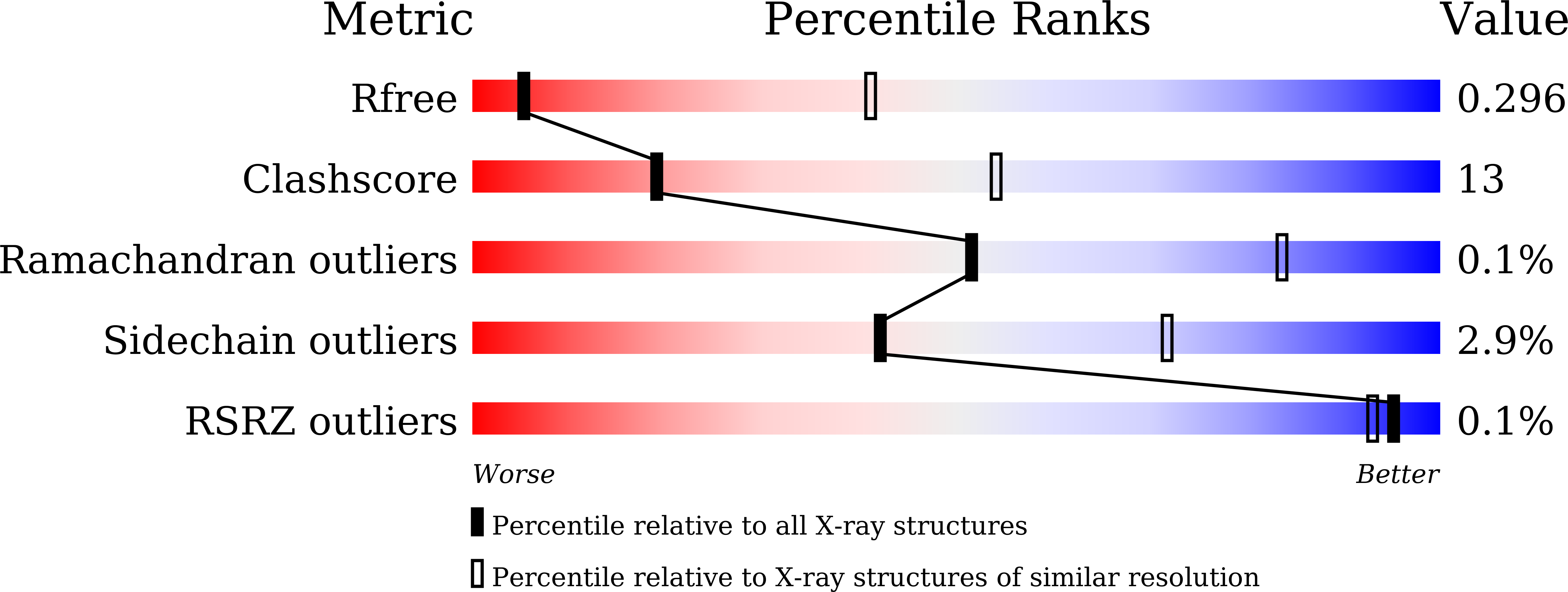
Experimental Data Snapshot
wwPDB Validation 3D Report Full Report
Entity ID: 1 | |||||
|---|---|---|---|---|---|
| Molecule | Chains | Sequence Length | Organism | Details | Image |
| Mycobacterium tuberculosis Mfd | 1,254 | Mycobacterium tuberculosis H37Rv | Mutation(s): 0 Gene Names: mfd EC: 3.6.4 | 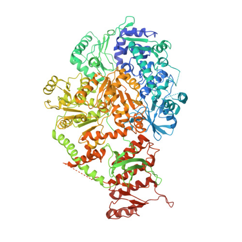 | |
UniProt | |||||
Find proteins for P9WMQ5 (Mycobacterium tuberculosis (strain ATCC 25618 / H37Rv)) Explore P9WMQ5 Go to UniProtKB: P9WMQ5 | |||||
Entity Groups | |||||
| Sequence Clusters | 30% Identity50% Identity70% Identity90% Identity95% Identity100% Identity | ||||
| UniProt Group | P9WMQ5 | ||||
Sequence AnnotationsExpand | |||||
| |||||
| Length ( Å ) | Angle ( ˚ ) |
|---|---|
| a = 224.259 | α = 90 |
| b = 224.259 | β = 90 |
| c = 224.259 | γ = 90 |
| Software Name | Purpose |
|---|---|
| PHENIX | refinement |
| iMOSFLM | data reduction |
| SCALA | data scaling |
| MOLREP | phasing |
| Funding Organization | Location | Grant Number |
|---|---|---|
| Other government | India | BT/HRD/35/02/19/2009 |
RCSB PDB (citation) is hosted by
RCSB PDB is a member of the

