6AAM

Crystal structure of TYK2 in complex with peficitinib


Experimental Data Snapshot

  • Method: X-RAY DIFFRACTION
  • Resolution: 1.98 Å
  • R-Value Free: 0.238 
  • R-Value Work: 0.202 
  • R-Value Observed: 0.204 

wwPDB Validation   3D Report Full Report


Ligand Structure Quality Assessment 


This is version 1.3 of the entry. See complete history


Literature

Discovery and structural characterization of peficitinib (ASP015K) as a novel and potent JAK inhibitor

Hamaguchi, H.Amano, Y.Moritomo, A.Shirakami, S.Nakajima, Y.Nakai, K.Nomura, N.Ito, M.Higashi, Y.Inoue, T.

(2018) Bioorg Med Chem 26: 4971-4983

  • DOI: https://doi.org/10.1016/j.bmc.2018.08.005
  • Primary Citation of Related Structures:  
    6AAH, 6AAJ, 6AAK, 6AAM

  • PubMed Abstract: 

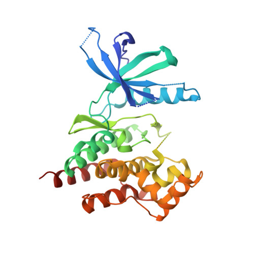
    Janus kinases (JAKs) are considered promising targets for the treatment of autoimmune diseases including rheumatoid arthritis (RA) due to their important role in multiple cytokine receptor signaling pathways. Recently, several JAK inhibitors have been developed for the treatment of RA. Here, we describe the identification of the novel orally bioavailable JAK inhibitor 18, peficitinib (also known as ASP015K), which showed moderate selectivity for JAK3 over JAK1, JAK2, and TYK2 in enzyme assays. Chemical modification at the C4-position of lead compound 5 led to a large increase in JAK inhibitory activity and metabolic stability in liver microsomes. Furthermore, we determined the crystal structures of JAK1, JAK2, JAK3, and TYK2 in a complex with peficitinib, and revealed that the 1H-pyrrolo[2,3-b]pyridine-5-carboxamide scaffold of peficitinib forms triple hydrogen bonds with the hinge region. Interestingly, the binding modes of peficitinib in the ATP-binding pockets differed among JAK1, JAK2, JAK3, and TYK2. WaterMap analysis of the crystal structures suggests that unfavorable water molecules are the likely reason for the difference in orientation of the 1H-pyrrolo[2,3-b]pyridine-5-carboxamide scaffold to the hinge region among JAKs.


  • Organizational Affiliation

    Drug Discovery Research, Astellas Pharma Inc., 21, Miyukigaoka, Tsukuba, Ibaraki 305-8585, Japan. Electronic address: hisao.hamaguchi@astellas.com.


Macromolecules
Find similar proteins by:  (by identity cutoff)  |  3D Structure
Entity ID: 1
MoleculeChains Sequence LengthOrganismDetailsImage
Non-receptor tyrosine-protein kinase TYK2298Homo sapiensMutation(s): 7 
Gene Names: TYK2
EC: 2.7.10.2
UniProt & NIH Common Fund Data Resources
Find proteins for P29597 (Homo sapiens)
Explore P29597 
Go to UniProtKB:  P29597
PHAROS:  P29597
GTEx:  ENSG00000105397 
Entity Groups  
Sequence Clusters30% Identity50% Identity70% Identity90% Identity95% Identity100% Identity
UniProt GroupP29597
Sequence Annotations
Expand
  • Reference Sequence
Small Molecules
Ligands 1 Unique
IDChains Name / Formula / InChI Key2D Diagram3D Interactions
9T6
Query on 9T6

Download Ideal Coordinates CCD File 
B [auth A]4-[[(1S,3R)-5-oxidanyl-2-adamantyl]amino]-1H-pyrrolo[2,3-b]pyridine-5-carboxamide
C18 H22 N4 O2
DREIJXJRTLTGJC-ZKVNVPQCSA-N
Binding Affinity Annotations 
IDSourceBinding Affinity
9T6 BindingDB:  6AAM IC50: min: 4.8, max: 5 (nM) from 2 assay(s)
Experimental Data & Validation

Experimental Data

  • Method: X-RAY DIFFRACTION
  • Resolution: 1.98 Å
  • R-Value Free: 0.238 
  • R-Value Work: 0.202 
  • R-Value Observed: 0.204 
  • Space Group: P 21 21 21
Unit Cell:
Length ( Å )Angle ( ˚ )
a = 36.142α = 90
b = 70.531β = 90
c = 97.891γ = 90
Software Package:
Software NamePurpose
REFMACrefinement
HKL-2000data reduction
HKL-2000data scaling
AMoREphasing

Structure Validation

View Full Validation Report


Ligand Structure Quality Assessment 


Entry History 

Deposition Data

Revision History  (Full details and data files)

  • Version 1.0: 2018-08-15
    Type: Initial release
  • Version 1.1: 2018-09-12
    Changes: Data collection, Database references
  • Version 1.2: 2018-10-24
    Changes: Data collection, Database references
  • Version 1.3: 2024-03-27
    Changes: Data collection, Database references