6A9C

Crystal Structure c-terminal SH3 domain of Myosin IB from Entamoeba histolytica bound to EhFP10(GEF) peptide.


Experimental Data Snapshot

  • Method: X-RAY DIFFRACTION
  • Resolution: 1.98 Å
  • R-Value Free: 0.240 
  • R-Value Work: 0.201 
  • R-Value Observed: 0.203 

wwPDB Validation   3D Report Full Report


This is version 1.1 of the entry. See complete history


Literature

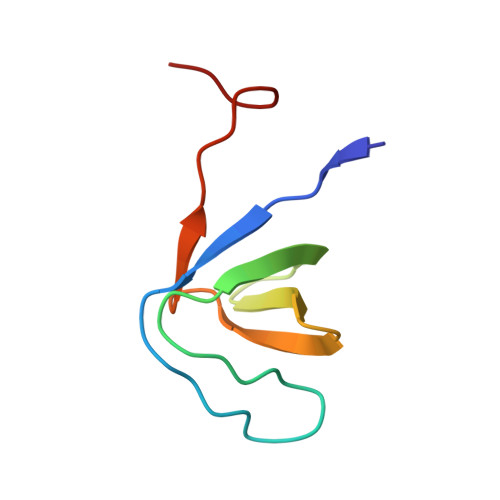
EhFP10: A FYVE family GEF interacts with myosin IB to regulate cytoskeletal dynamics during endocytosis in Entamoeba histolytica.

Gautam, G.Ali, M.S.Bhattacharya, A.Gourinath, S.

(2019) PLoS Pathog 15: e1007573-e1007573

  • DOI: https://doi.org/10.1371/journal.ppat.1007573
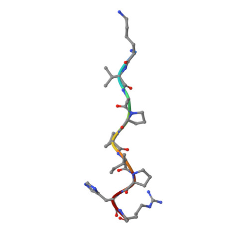
  • Primary Citation of Related Structures:  
    6A9C

  • PubMed Abstract: 

    Motility and phagocytosis are key processes that are involved in invasive amoebiasis disease caused by intestinal parasite Entamoeba histolytica. Previous studies have reported unconventional myosins to play significant role in membrane based motility as well as endocytic processes. EhMyosin IB is the only unconventional myosin present in E. histolytica, is thought to be involved in both of these processes. Here, we report an interaction between the SH3 domain of EhMyosin IB and c-terminal domain of EhFP10, a Rho guanine nucleotide exchange factor. EhFP10 was found to be confined to Entamoeba species only, and to contain a c-terminal domain that binds and bundles actin filaments. EhFP10 was observed to localize in the membrane ruffles, phagocytic and macropinocytic cups of E. histolytica trophozoites. It was also found in early pinosomes but not early phagosomes. A crystal structure of the c-terminal SH3 domain of EhMyosin IB (EhMySH3) in complex with an EhFP10 peptide and co-localization studies established the interaction of EhMySH3 with EhFP10. This interaction was shown to lead to inhibition of actin bundling activity and to thereby regulate actin dynamics during endocytosis. We hypothesize that unique domain architecture of EhFP10 might be compensating the absence of Wasp and related proteins in Entamoeba, which are known partners of myosin SH3 domains in other eukaryotes. Our findings also highlights the role of actin bundling during endocytosis.


  • Organizational Affiliation

    School of Life Sciences, Jawaharlal Nehru University, New Delhi, India.


Macromolecules
Find similar proteins by:  (by identity cutoff)  |  3D Structure
Entity ID: 1
MoleculeChains Sequence LengthOrganismDetailsImage
Unconventional myosin IBA [auth B],
B [auth A]
66Entamoeba histolyticaMutation(s): 0 
Gene Names: EHI_110810
UniProt
Find proteins for C4LUC7 (Entamoeba histolytica (strain ATCC 30459 / HM-1:IMSS / ABRM))
Explore C4LUC7 
Go to UniProtKB:  C4LUC7
Entity Groups  
Sequence Clusters30% Identity50% Identity70% Identity90% Identity95% Identity100% Identity
UniProt GroupC4LUC7
Sequence Annotations
Expand
  • Reference Sequence

Find similar proteins by:  Sequence   |   3D Structure  

Entity ID: 2
MoleculeChains Sequence LengthOrganismDetailsImage
Peptide from Rho guanine nucleotide exchange factorC [auth E]9Entamoeba histolyticaMutation(s): 0 
UniProt
Find proteins for C4M4E9 (Entamoeba histolytica (strain ATCC 30459 / HM-1:IMSS / ABRM))
Explore C4M4E9 
Go to UniProtKB:  C4M4E9
Entity Groups  
UniProt GroupC4M4E9
Sequence Annotations
Expand
  • Reference Sequence
Experimental Data & Validation

Experimental Data

  • Method: X-RAY DIFFRACTION
  • Resolution: 1.98 Å
  • R-Value Free: 0.240 
  • R-Value Work: 0.201 
  • R-Value Observed: 0.203 
  • Space Group: P 21 21 21
Unit Cell:
Length ( Å )Angle ( ˚ )
a = 29.008α = 90
b = 60.013β = 90
c = 95.305γ = 90
Software Package:
Software NamePurpose
REFMACrefinement
HKL-2000data reduction
HKL-2000data scaling
PHASERphasing

Structure Validation

View Full Validation Report


Entry History 

Deposition Data

Revision History  (Full details and data files)

  • Version 1.0: 2019-06-12
    Type: Initial release
  • Version 1.1: 2023-11-22
    Changes: Data collection, Database references, Refinement description