6A48

Crystal structure of reelin N-terminal region


Experimental Data Snapshot

  • Method: X-RAY DIFFRACTION
  • Resolution: 2.00 Å
  • R-Value Free: 0.257 
  • R-Value Work: 0.215 
  • R-Value Observed: 0.217 

wwPDB Validation   3D Report Full Report


Ligand Structure Quality Assessment 


This is version 1.3 of the entry. See complete history


Literature

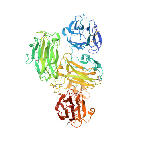
Structural studies of reelin N-terminal region provides insights into a unique structural arrangement and functional multimerization.

Nagae, M.Suzuki, K.Yasui, N.Nogi, T.Kohno, T.Hattori, M.Takagi, J.

(2021) J Biochem 169: 555-564

  • DOI: https://doi.org/10.1093/jb/mvaa144
  • Primary Citation of Related Structures:  
    6A48

  • PubMed Abstract: 

    The large, secreted glycoprotein reelin regulates embryonic brain development as well as adult brain functions. Although reelin binds to its receptors via its central part, the N-terminal region directs multimer formation and is critical for efficient signal transduction. In fact, the inhibitory antibody CR-50 interacts with the N-terminal region and prevents higher-order multimerization and signalling. Reelin is a multidomain protein in which the central part is composed of eight characteristic repeats, named reelin repeats, each of which is further divided by insertion of a epidermal growth factor (EGF) module into two subrepeats. In contrast, the N-terminal region shows unique 'irregular' domain architecture since it comprises three consecutive subrepeats without the intervening EGF module. Here, we determined the crystal structure of the murine reelin fragment named RX-R1 including the irregular region and the first reelin repeat at 2.0-Å resolution. The overall structure of RX-R1 has a branched Y-shaped form. Interestingly, two incomplete subrepeats cooperatively form one entire subrepeat structure, though an additional subrepeat is inserted between them. We further reveal that Arg335 of RX-R1 is crucial for binding CR-50. A possible self-association mechanism via the N-terminal region is proposed based on our results.


  • Organizational Affiliation

    Department of Molecular Immunology, Research Institute for Microbial Diseases.


Macromolecules
Find similar proteins by:  (by identity cutoff)  |  3D Structure
Entity ID: 1
MoleculeChains Sequence LengthOrganismDetailsImage
Reelin677Mus musculusMutation(s): 0 
Gene Names: RelnRl
EC: 3.4.21
UniProt & NIH Common Fund Data Resources
Find proteins for Q60841 (Mus musculus)
Explore Q60841 
Go to UniProtKB:  Q60841
IMPC:  MGI:103022
Entity Groups  
Sequence Clusters30% Identity50% Identity70% Identity90% Identity95% Identity100% Identity
UniProt GroupQ60841
Sequence Annotations
Expand
  • Reference Sequence
Experimental Data & Validation

Experimental Data

  • Method: X-RAY DIFFRACTION
  • Resolution: 2.00 Å
  • R-Value Free: 0.257 
  • R-Value Work: 0.215 
  • R-Value Observed: 0.217 
  • Space Group: P 21 21 21
Unit Cell:
Length ( Å )Angle ( ˚ )
a = 84.753α = 90
b = 90.999β = 90
c = 90.535γ = 90
Software Package:
Software NamePurpose
PHENIXrefinement
HKL-2000data reduction
HKL-2000data scaling
MOLREPphasing

Structure Validation

View Full Validation Report


Ligand Structure Quality Assessment 


Entry History 

Deposition Data

Revision History  (Full details and data files)

  • Version 1.0: 2019-06-19
    Type: Initial release
  • Version 1.1: 2020-07-29
    Type: Remediation
    Reason: Carbohydrate remediation
    Changes: Data collection, Derived calculations, Structure summary
  • Version 1.2: 2022-06-29
    Changes: Database references, Structure summary
  • Version 1.3: 2023-11-22
    Changes: Data collection, Refinement description